
Stratified squamous epithelium
Various layers of overlapping cells form this epithelium. The side of these cells near the free margin of the tissue appears flattened. It has a protective, barrier function against external aggressions. It can either be keratinized or non-keratinized (the latter type is actually partially keratinized).

Figure E59. Schematic drawing of the organization of the stratified squamous epithelium.
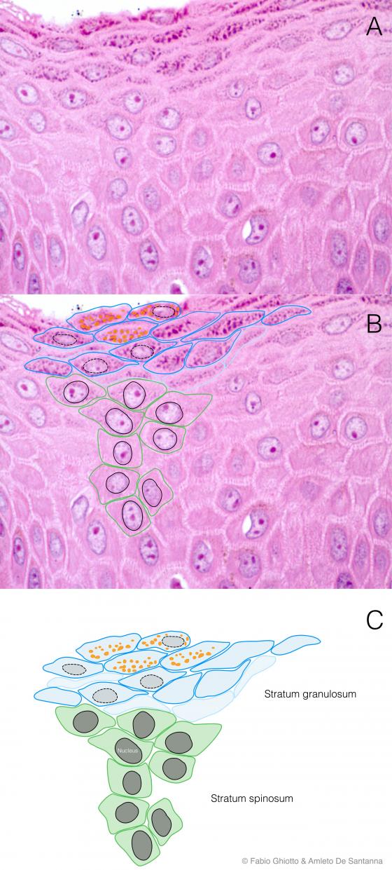
Figure E60. Digitally annotated micrograph of the non-keratinized stratified squamous epithelium. Epithelial cells are flattened and located on several layers. The number of layers is variable, depending on the amount of wear the epithelium is subject to (more wear, more cell layers). The definition of squamous epithelium (as for all stratified tissues) is due to the shape of the cells in the more superficial layers.
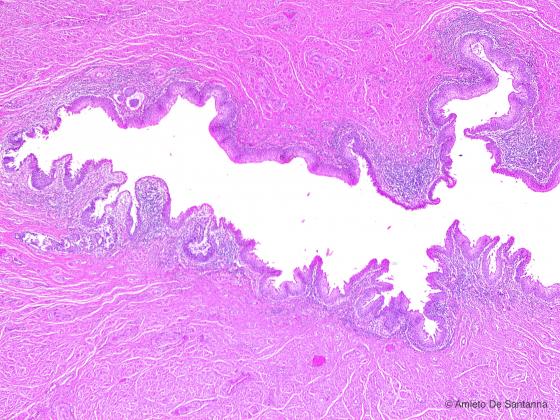
Figure E61. Human female urethra. Stratified squamous epithelium of the female urethra. Differently from the male urethra, the short female urethra is lined only by this type of epithelium. H&E X25
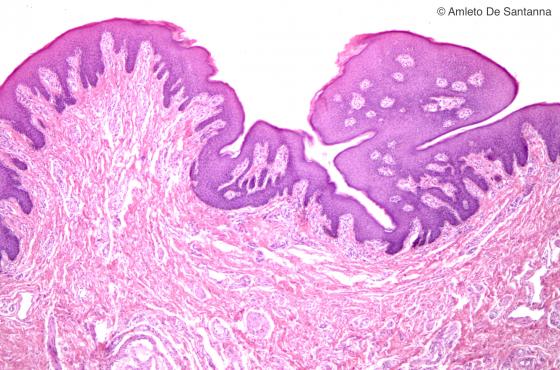
Figure E62. Human prepuce. Stratified squamous epithelium. H&E X40
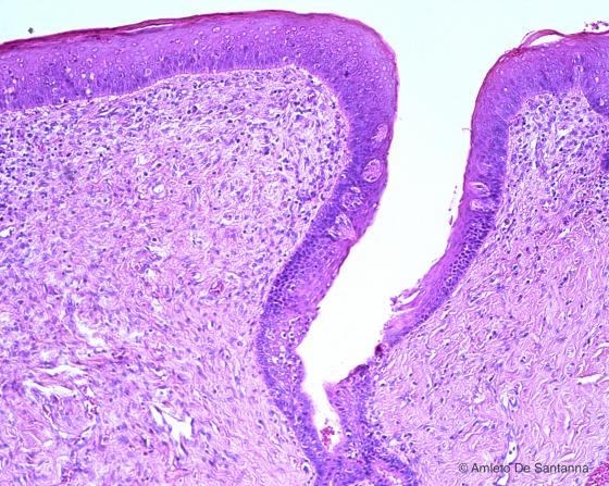
Figure E63. Human fetal tongue. Circumvallate papillae lined by stratified squamous epithelium. H&E X100
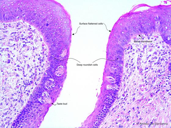
Figure E64. Human fetal tongue. Circumvallate papillae at higher magnification. You can see the morphological difference of the cells that compose the different layers of the stratified squamous epithelium. The superficial layer(s) is (are) made of highly flattened cells, whereas, in the layers below, cells have a cubical or columnar shape. To categorize the stratified epithelia you must always take into account the cell shape of the superficial layers. H&E X200
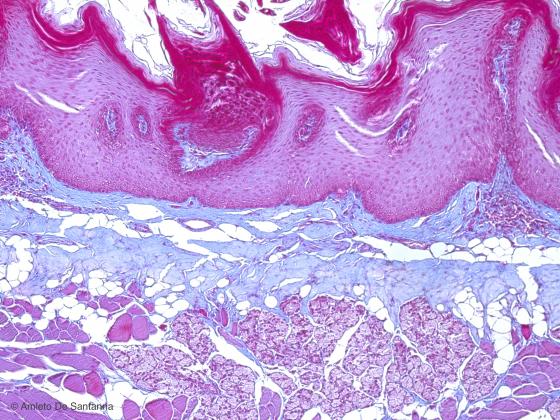
Figure E65. Human fetal tongue. Lingual mucosa lined by stratified squamous epithelium. Mallory-Azan X100
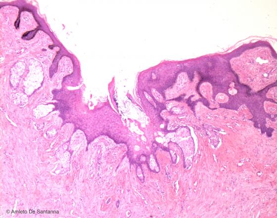
Figure E66. Human mammary papilla (nipple). Stratified squamous epithelium. H&E X40
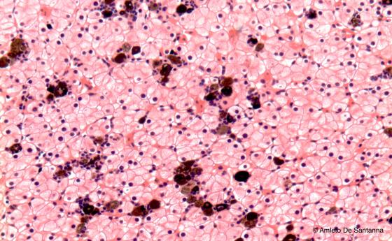
Figure E67. Human skin. Transversal section of pigmented human skin. H&E X40
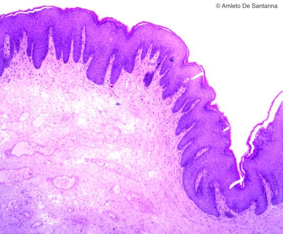
Figure E68. Human anus. Stratified squamous epithelium lining the anal canal. H&E X40
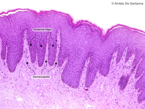
Figure E69. Human anus. Stratified squamous epithelium lining the anal canal. You can easily see the dermal papillae and the epidermal ridges. This arrangement allows a better anchoring of the epidermis on the underlying dermis. H&E X100
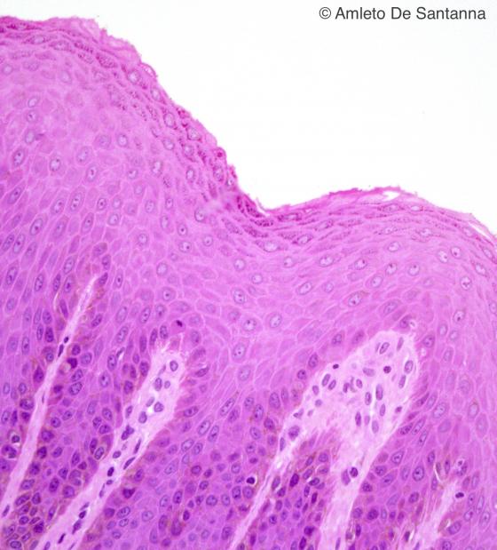
Figure E70. Human anus. Higher magnification of the stratified squamous epithelium lining the anal canal. You can easily see the following layers: the stratum spinosum and the stratum granulosum. H&E X250
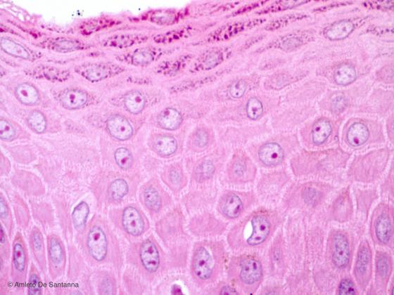
Figure E71. Human anus. High magnification micrograph of the stratified squamous epithelium lining the anal canal. You can easily see the following layers: the stratum spinosum and the stratum granulosum. H&E X630
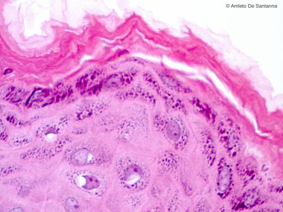
Figure E72. Human skin at high magnification. You can see the stratum granulosum and the stratum corneum. H&E X630

Figure E73. Human anorectal junction. Region of the large intestine where the simple columnar epithelium of the rectum (right) is replaced (arrow) by the stratified squamous epithelium of the anus (left). H&E X63

Figure E74. Mouse esophagus. Stratified squamous epithelium. You can see the different layers of epithelial cells that are cuboidal in the deepest layer and become gradually more flattened as you move to the surface. Ignesti X100

Figure E75. Human palmar skin. Highly keratinized stratified squamous epithelium (thick skin). Flattened cells are arranged in several layers and, on the surface of the epithelium, there is a thick stratum corneum. H&E X63
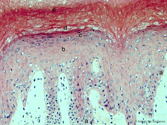
Figure E76. Human skin. From the dermal ridges, some elements are clearly visible: a) the stratum basale (stratum germinativum), made of a single layer of cuboidal or columnar cells; b) the stratum spinosum (spinous layer), so called because of its several layers of cuboidal or flattened cells having cytoplasmic processes that resemble spines; c) the stratum granulosum, made of layers of flattened cells which show keratohyalin granules; d) the stratum lucidum, made of squamous cells without nucleus and therefore colorless.This layer sometimes is not present; e) the stratum corneum, made of a several layers of squamous cells without nucleus and completely keratinized. H&E X100
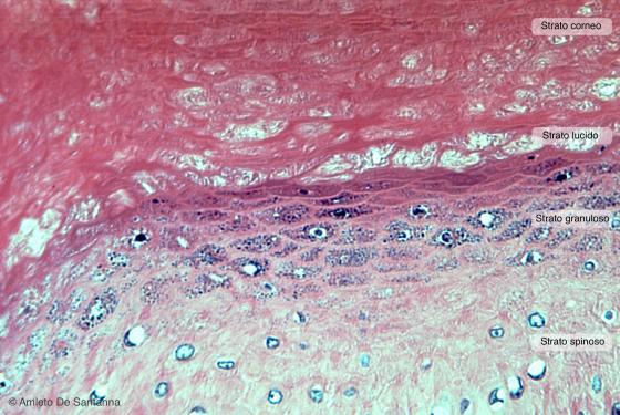
Figure E77. Squamous stratified epithelium. Stratum spinosus, stratum garnulosum, stratum lucidum and stratum spinosus at high magnification. You can see that, at this magnification power, the cytoplasmic processes of the stratum spinosus are not easily visible. In order to make them visible you need a specific staining method for cellular borders like iron hematoxylin. H&E X200
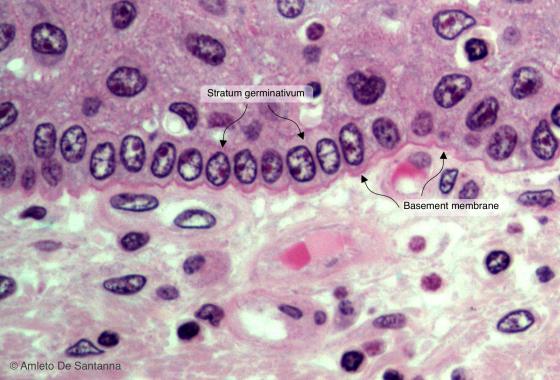
Figure E78. Human skin. Stratified squamous epithelium. Stratum basale (stratum germinativum) at high magnification. H&E X630
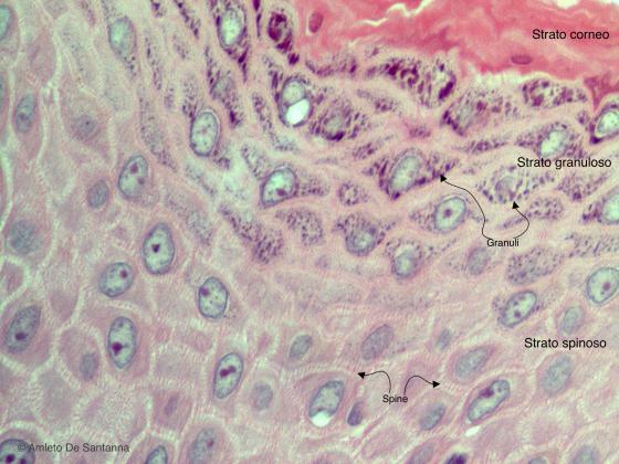
Figure E79. Human skin. Stratified squamous epithelium at high magnification where you can see the cytoplasmic processes (spines) and the keratohyalin granules. H&E X630
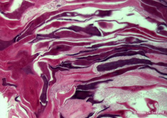
Figure E80. Human skin. Keratin layers in the keratinized stratified squamous epithelium. H&E X630
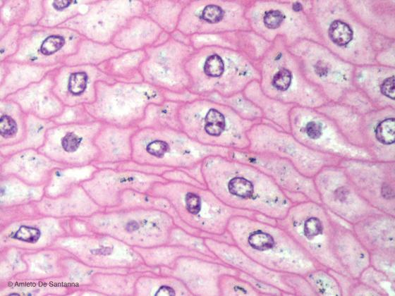
Figure E81. Human skin. Stratified squamous epithelium in transversalsection. The cellular borders are clearly visible. H&E X630
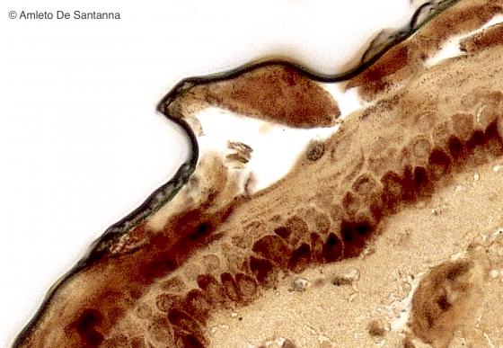
Figure E82. Human skin. Sample from a black skinned person where you can see the large amount of melanin in the epidermis. Iron hematoxylin X400
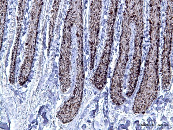
Figure E83. Whale skin. Melanin granules of a marine mammal. Hematoxylin-DAB X100
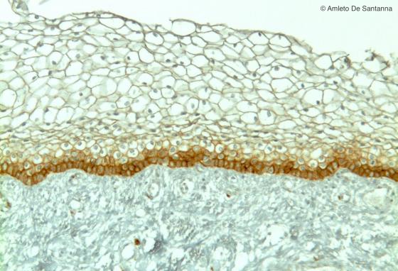
Figure E84. Human fetus. Stratified squamous epithelium of a portion of the nose. Immunohistochemical reaction using a monoclonal antibody then recognizes the CD156 antigen expressed by the cells of the germinal layer of the stratified epithelia. The cells of the germinal layer are stained in deep brown. EM-DAB X100
Stratified cuboidal epithelium
Two or more layers of cuboidal cells compose the stratified cuboidal epithelium. In mammals, this epithelium is very rare and covers only some large excretory duct.
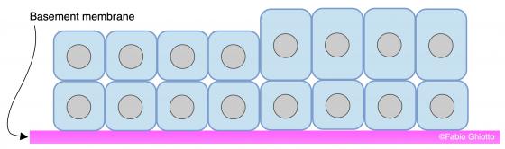
Figure E85. Schematic drawing of the stratified cuboidal epithelium.
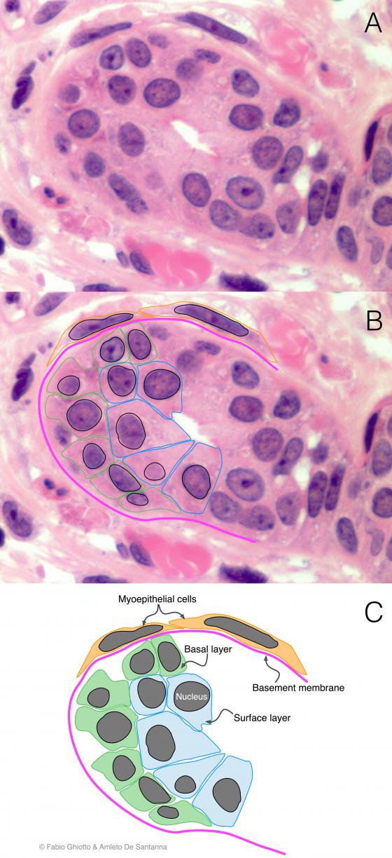
Figure E86. Digitally annotated micrograph of stratified cuboidal epithelium. The cells of the cuboidal epithelia have approximately the same width, depth and height and their nuclei are roundish and positioned centrally. The stratified cuboidal epithelium is made by no more than two/three layers (colored differently in the figure). You can see, outside the excretory duct, two myoepithelial cells (orange).
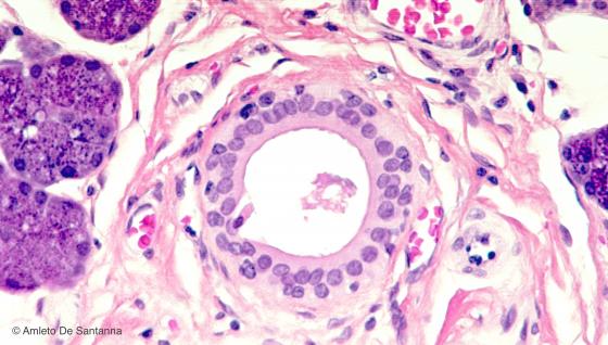
Figure E87. Human submandibular gland. Excretory duct lined by stratified cuboidal epithelium. The number of the cell layers, in this type of epithelium, is always low, two or three layers at most. H&E X200
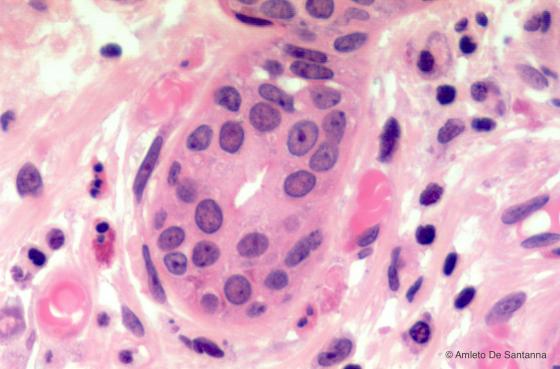
Figure E88. Human parotid gland. Excretory duct lined by stratified cuboidal epithelium. H&E X400
Stratified columnar epithelium
Two or more layers of overlapping cells compose this epithelium. The ones in the deepest layer are smaller, polyhedral and they never reach the surface of the epithelium, while the most superficial layer presents cells with a fully-fledged columnar shape. In tissue specimens, nuclei seem to be overlapping. The function of this epithelium is to protect and cover large ducts or highly sprinkled surfaces, and it is not easily found in mammals. It can only be found in large excretory ducts, in the penile urethra, in a short tract of the epiglottis and on the inner surface of the eyelid.
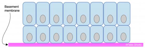
Figure E89. Schematic drawing of the stratified columnar epithelium.
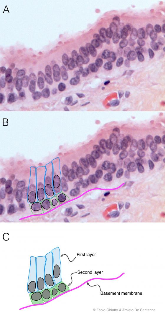
Figure E90. Digitally annotated micrograph of stratified columnar epithelium. The columnar cells are neatly superimposed in two (three) layers. This arrangement clearly differentiates this epithelium from the pseudostratified epithelium. You can see that the columnar cells are in the most external layer.
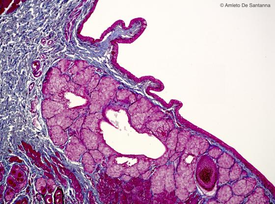
Figure E91. Human eyelid. The stratified columnar epithelium is infrequent. We find it when the epithelial tissue needs to contain strong fluxes of biological fluids or secretions. An example is the internal side of the eyelid. Mallory-Azan X40
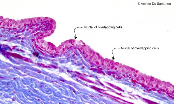
Figure E92. Human eyelid. You can see the cells that, overlapping on each other, make up the different layers of the stratified columnar epithelium that lines the internal surface of the eyelid (conjunctiva). Mallory-Azan X250
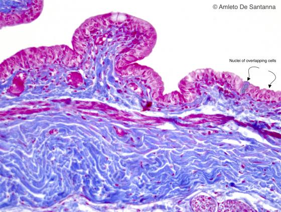
Figure E93. Human eyelid. Stratified columnar epithelium at higher magnification. Mallory-AzanX400
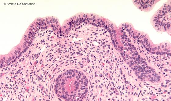
Figure E94. Human penile urethra. Short segment of the urethra lined by stratified columnar epithelium. The deepest layer of the epithelium is made by small cuboidal cells. Differently, the superficial layer is made by tall columnar cells. You can distinguish the stratified columnar epithelium from the pseudostratified epithelium because in the first you have at least two superimposed cells (one over the other) whereas in the pseudostratified epithelium, the nuclei seem to be on different layers but never superimposed. H&E X100
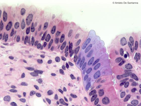
Figure E95. Human penile urethra. Stratified columnar epithelium at high magnification. The favorable section plane let you see clearly the layer arrangement of the epithelial cells (shaded area). H&E X400
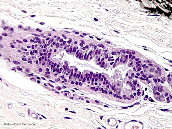
Figure E96. Human breast. Stratified columnar epithelium lining a duct of a human breast gland. H&E X250
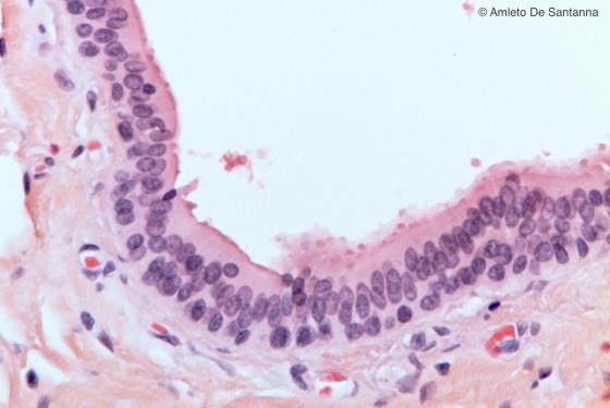
Figure E97. Human submandibular gland. Stratified columnar epithelium lining a large excretory duct. H&E X200
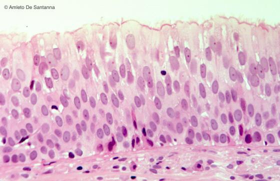
Figure E98. Human larynx. This is the only case where we find a ciliated stratified columnar epithelium in a human tissue. H&E X200
Transitional epithelium
The morphology of this epithelium presents much variability, because it covers organs such as the urinary bladder and the ureter, whose volume varies a lot. Three types of cells compose this epithelium: the basal layer, in direct contact with the basement membrane, composed of cuboidal or columnar cells (stratum basale or germinativum). Above them, some intermediate layers of elongated cells (also known as clavate or pear-shaped) are found. Finally, the superficial layer, whose cells may sometimes be binucleated. The long axis of these cells is orthogonal with respect to the long cell axis of the intermediate layer and their top surface has a convex shape; this explains why they are known as dome-shaped or umbrella cells. This epithelium can increase its surface following the dilatation of organs, since the cells of the intermediate layer can deform and stretch on a horizontal plane and still remain stuck in the umbrella cells.
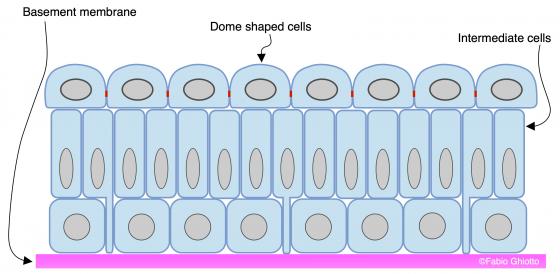
Figure E99. Schematic drawing of the transitional epithelium (urothelium) (according to S.P. Jost, J.A. Gosling & J.S. Dixon: The morphology of normal human bladder urothelium. J. Anat., 1989, 167 pp. 103-115).
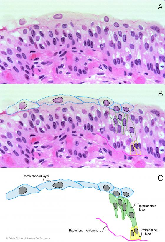
Figure E100. Digitally annotated micrograph of transitional epithelium. This epithelium is made up of cells that have a different morphology, depending on the layer they belong to. The epithelium is made of a basal cell layer where the cells are mainly cuboidal (yellow), intermediate cell layers where cells are tall (green) and a superficial dome-shaped cell layer (light blue), where cells sometimes have two nuclei. This arrangement makes the tissue suitable for remarkable stretching.
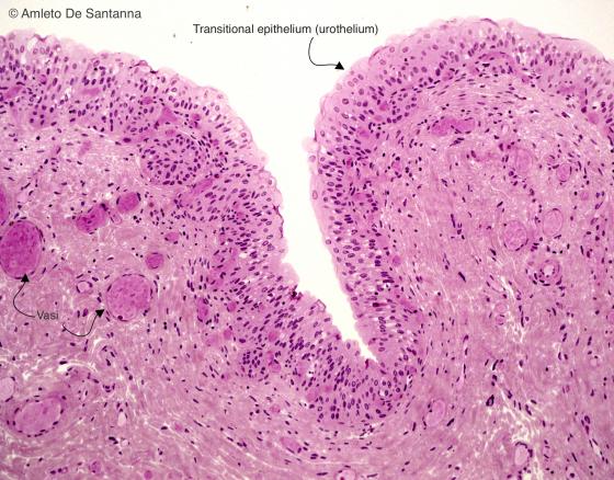
Figure E101. Human urinary bladder. Transitional epithelium at low magnification. You can see that there is no submucosa. H&E X40
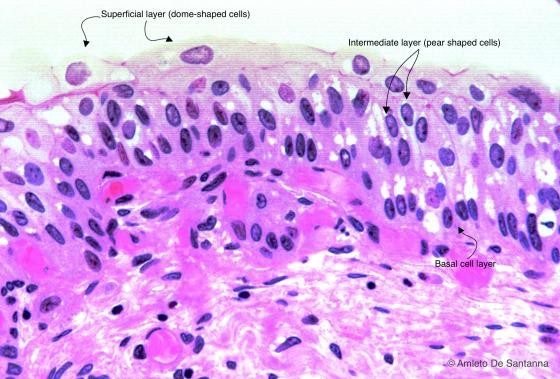
Figure E102. Human urinary bladder. Transitional epithelium at high magnification. A single layer of cuboidal or columnar cells lies on the basement membrane. On top of this layer, there are several layers of tall cells (intermediate cells). The most superficial layer is made of dome shaped cells, sometimes binucleated. H&E X200

Figure E103. Human ureter. Transitional epithelium. H&E X63
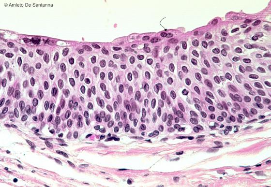
Figure E104. Human ureter. Transitional epithelium. In this micrograph, you can easily see the different cell layers and, on the surface, a dome shaped binucleated cell (arrow). H&E X200