
The term connective tissue describes a type of tissue that supports other tissues, both structurally and metabolically. This is the reason why it is also known as “supportive tissue”.
All different types of connective tissue originate from mesenchyme. Mesoderm-derived mesenchymal cells present an irregular, generally elongated shape and an amorphous extracellular substance, which is free of fibers. These are pluripotent cells, which means they can differentiate into muscle cells and the various connective tissue cell types: fibroblasts, chondrocytes, adipocytes, erythrocytes (red blood cells or RBCs), leukocytes (white blood cells or WBCs), macrophages.
The composition of connective tissue includes its cells and a large amount of extracellular matrix. The nature of the matrix determines the characteristics of each connective tissue type. There are liquid extracellular matrixes, such as in blood and lymph, through which different types of connective cells can be transported (erythrocytes, leukocytes, etc.). There are solid but loose extracellular matrixes that allow the passage of blood vessels and substances. There are also calcified matrixes (bone tissue) which create highly resistant tissues. Both the type of cells and the nature of the extracellular matrix distinguish the various types of connective tissue. The matrix is also called “extracellular substance” and its structure is made of two components: fibers (made of collagen and other types of proteins) and the amorphous ground substance (made of proteoglycans). The fiber nature, morphology and texture vary widely, depending on the type of connective tissue and on its functions, on the age of the individual and the specific characteristics of the sample species.
The varied structure of the connective tissue matches its functions. For example, loose connective tissue, due to its light, highly vascularized structure, easy to penetrate and full of blood and lymph vessels, represents the adequate solution for trophic function, transport of useful substances and protection against viral and bacterial infections.
Conversely, for containment, support or barrier function (as for skin) we need a dense connective tissue. Its structure is dense, very compact and presents a large amount of fibers, properly organized and oriented in order to comply with their functions.
In addition, fiber organization and nature vary depending on the function of the connective tissue analyzed. Underneath every simple or stratified epithelium, there is a loose connective tissue packed with reticular fibers that supports the epithelium and allows substances to spread from capillaries to epithelial cells. In case the connective tissue needs to resist stretch and distension, as tendons do, it must be packed with adequately disposed collagen and elastic fibers. In case the tissue needs to resist compression or surrounds a large organ, then collagen will be the main component of connective fibers.
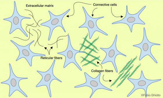
Figure C1. Schematic drawing showing the organization of the connective tissue.
Collagen fibers
Fibroblasts secrete collagen fibers that can be found in the majority of connective tissues. A family of proteins secreted as procollagen, forms them. Procollagen is then processed to tropocollagen in an extracellular environment. The tropocollagen creates fibers, different in quantity, that may appear as thin strands as well as thick, interwoven fibers (up to 10 µm). Their length is not easily determined, as they spread in different directions and, depending on their functions, they are disposed into parallel, interwoven or crossed rows. In case they are not stretched, their pattern looks slightly wavy.
Collagen fibers take on a pink color with Em-Eo, and a bluish color with Mallory. There are specific color techniques for reticular (type III collagen) and elastic fibers. For this type of tissue, intensity is proportional to the number and thickness of the fibers contained. This is the reason why dense connective tissue will always appear more colorful than loose connective tissue.
| Type | Distribution | Organization |
|---|---|---|
| I | bone, tendons, dentin, skin | thick parallel or interwoven fibers with evident periodic bands |
| II | ialine and elastic cartilage, vitreous body | thin fibers |
| III | reticular connective tissue, loose connective tissue coating the muscle fibers | thin fibers that coat other tissue or that make the weave of several organs |
| IV | basement membranes | fibers that are under the epithelia, thin and interwoven to form a network |
| V | amniotic sac, chorion, muscle and tendon sheaths | it doesn't form bands |
Table C1. Main types of collagen.
Elastic fibers
Fibroblasts, chondroblasts, chondrocytes (and smooth muscle cells in muscle tissue) synthesize these fibers. Their main component is elastin; in fact, in many organs it is important that the connective tissue is elastic. Elastic fibers can slide, stretch or deform, due to external impulses. In this way, they allow organ extension and contraction and have the ability to make organs or tissues return to their original shape. A connective tissue rich in elastic fibers can be found in the epiglottis, the urinary bladder and the tunica media of arteries. In ordinary, topographic techniques, such as Em-Eo or Mallory-Azan, the thin elastic fibers are not easily distinguished from the other fibers. However, while they rest and do not stretch, they can be distinguished by their distinctly wavy pattern. For a meticulous research of elastic fibers presence, selective dyes such as Weigert’s resorcin-fuchsin must be used. On an almost colorless background, this dye makes elastin pop with an intense purple-black.
Reticular fibers
They compose both the connective meshwork (stroma), whose function is to support the parenchyma of larger endocrine and exocrine glands, and the meshwork surrounding adipose and nerve cells, as well as muscle fibers. They are connected to the epithelial basal laminae and support hematopoietic and lymphatic organs.
As elastic fibers, also reticular fibers are not easily distinguished if conventional dyes are used. In order to visualize them, it is necessary to exploit their ability to bind metallic silver: to do so, alkaline solutions of silver salts are recommended. There are several specific metallic silver dyes for reticular fibers; one of the most popular is Bielschowsky’s.
Amorphous ground substance
The amorphous ground substance or extracellular amorphous ground substance is a viscous, colloidal liquid, which embeds connective tissue cells and fibers. This substance is a three-dimensional network, which can be more or less dense, formed by glycosaminoglycans (GAGs), glycoproteins and proteoglycan clusters. Their function is to bind water and make the matrix more permeable to metabolic substances and gasses that pass from blood to cells and vice versa. Therefore, amorphous matrix and tissue liquids are closely linked and perform the typical trophic function of loose connective tissue. This substance performs various functions: it links different parts of fiber structures in the most appropriate way, regulates the spread of metabolic substances and protects the organism by impeding the passage of harmful substances and pathogens. With aging, the extracellular substance of connective tissue undergoes major alterations.
During histological preparations, it dissolves and therefore it is impossible to find it stained inside the specimens. Inside a cryostat, if it is sectioned at -30°C and stained with PAS reaction or basic dyes, the result will still be mild. In cartilage and bone tissue, the abundance of glycosaminoglycans (GAGs) and the texture of the amorphous matrix ensure a good resistance to fixatives and chemical solvents. This allows for a good result with alcian blue, since this dye can bind chondroitin sulfates (a type of GAGs). Iron hematoxylin (as a histological dye) and alcian-PAS (as a histochemical dye) are usually employed to show the ground substance. Iron hematoxylin non-selectively highlights the extracellular substance between epithelial cells or connective fibers with an intense black dye. Alcian-PAS is a double histochemical dye for glycosaminoglycans. Alcian highlights acid glycosaminoglycans with an intense blue dye, while PAS, by the Schiff reagent, reveals neutral glycosaminoglycans with a pink-red one. Mature hyaline cartilage takes on a very intense shade with alcian blue, due to the presence of the highly acid chondroitin sulfate and heparan sulfate.
There are three main categories of connective tissue:
- Embryonic connective tissue
- Connective tissue proper
- Specialized connective tissue
Note: different texts might contain a different classification of connective tissue; for example, in some texts, the adipose tissue might be classified as connective tissue proper.