
This is a complex, highly-specific method used to reveal antigens present in the tissue or in the cells to analyze.
We need to place the specific antibody for the antigen we are looking for on a section of tissue or cells adequately prepared. In this way, we will get an immune antigen-antibody reaction. This reaction will be subsequently revealed by a secondary antibody conjugated to a catalysing enzyme (e.g. peroxidase). This enzyme reacts with a substrate (chromogenic, e.g. DAB, FAST RED-TR) and develops an insoluble, stained precipitate visible with a light microscope.
Depending on the available material and on the result we want to obtain, various methods may be used:
- Direct method. We take a specific antibody conjugated to the peroxidase (catalysing enzyme) and once it has been placed on the tissue, it will specifically conjugate to the antigen and allow the revelation through a chromogenic substrate (brown DAB, red FAST-RED, blue BPIC). The direct immunohistochemical method is generally very quick and non-specific reactions are unlikely to happen. The main problem is that for each antigen we aim to localize, a different conjugated antibody is needed. This method is traditionally used to detect complementary immunoglobulins and immune complexes present in kidney and muscle histological specimens from patients suffering from Systemic Lupus Erythematosus (SLE).
- Indirect method. A primary antibody sampled from an animal (rat, mouse, rabbit) reacts with the antigen which is possibly present on a tissue section. Subsequently, a conjugated secondary antibody, specific for the antibodies belonging to the animal previously used, is added; eventually, such antibodies are revealed using a stained substrate.
On the one hand, the direct method includes a one-to-one relationship between the catalyst and the antigen, and it develops a good amount of stained substrate by the end of the reaction. On the other hand, the indirect method presents a different relationship between the catalyst and the antigen (up to one-to-sixteen), since several secondary and tertiary antibodies bind to the primary antibody (such procedure is known as “signal amplification”). In this way, a greater amount of catalysts will bind to the antigen and develop a higher quantity of stained substrate by the end of the reaction, so that the antigen will appear clearer.
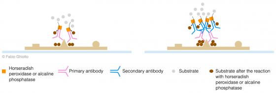
Figure S31. Schematic example of direct (on the left) and indirect (on the right) immuno-histochemical reactions.
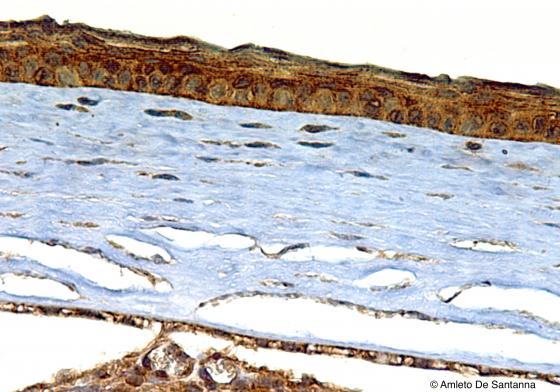
Figure S32. Mouse cornea. HRP-streptavidin-biotin immunohistochemical reaction that stains e-cadherins, with DAB, deep brown. X200
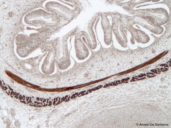
Figure S33. Fetal human intestine. HRP-streptavidin-biotin immunohistochemical reaction that highlights deep brown the forming smooth muscle fascicles. X63