
They provide information about the content and the nature of the chemical substances included into the biological tissue analyzed, rather than on the morphological aspect of the specimen. Therefore, one or more chemical reactions are performed. These operations are stoichiometrically exact and specific, and they highlight a determined functional group, or a specific ion present in the different tissues and in the cells which are part of them. “Sets of reactions” are performed in order to get, not only morphological evidence, but answers that will have to be incontrovertible and unique in the end. The stained precipitates obtained in this way will be visible with the light microscope or, in case they are fluorochromes, with the fluorescence microscope.
McManus-Hotchkiss (PAS reaction)
This reaction is specific for detecting neutral mucins in the tissues, such as the secreting epithelia, the endocrine glands, the nervous system. An oxidant is used (in this case the periodic acid) and it selectively attacks the primary amine functional group or the -OH one, releasing an aldehyde group which is subsequently revealed by the Schiff’s reagent (purple red). It is generally performed together with alcian blue.
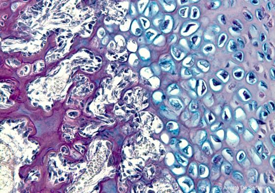
Figure S27. Inner ear of human embryo. Indirect (endochondral) ossification. The bone is stained magenta because of the high amount of neutral glycosaminoglycans. Cartilage, here in degeneration, is stained blue because of the high amount of acidic glycosaminoglycans. Alcian PAS X100
Alcian blue
It is an important reaction that highlights qualitatively and quantitatively both the sulfur acid mucins and the glycosaminoglycans (GAGs) containing carboxylic groups. It is usually performed simultaneously with the PAS reaction, in order to get a complete visualization of the specimen’s mucins, both acidic (Alcian, intense blue precipitate) and neutral (PAS, red). If the reaction takes place in a highly acidic environment (pH 1), then only the sulfur mucopolysaccharides will be marked. If instead it takes place in an average acid environment (pH 3), then also the carboxylic mucopolysaccharides will be marked.
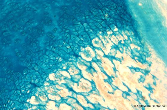
Figure S28. Indirect (endochondral) ossification in a human embryo. Cartilage is stained deep blue because of the large content of acidic glycosaminoglycans. Alcian blue X63
Feulgen reaction
This is a histochemical reaction which stains the DNA magenta. The hydrolysis takes place in an oven at 60°C with HCl 1N and its duration is variable: it depends on the fixatives previously used, on the tissue and on the animal. This is the most delicate phase of the reaction, because the purinic bases separate with hot hydrolysis and aldehyde groups develop, and become stained magenta by the Schiff’s reagent. In case the hydrolysis is prolonged longer than recommended, even histones and apurinic acids are distanced. In this way, the reaction of the hydrolysed liquid increases and chromosomes will appear less vivid. This reaction is highly specific, since there is no other known cellular substance that, in these conditions, can develop a stained compost with the Schiff’s reagent.
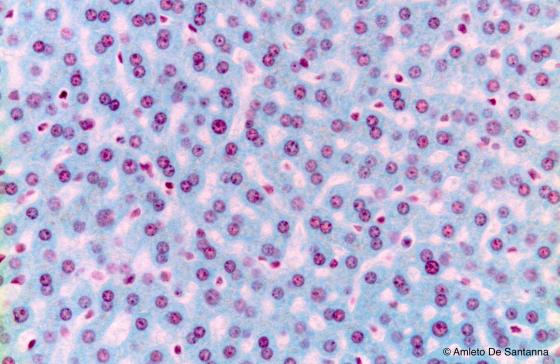
Figure S29. Mouse liver. Feulgen reaction for DNA unmasking. Feulgen X100
Hillarp reaction
Hillarp reaction for chromaffin cells is a specific reaction for the catecholamines found, for example, in the suprarenal medulla cells (secreting adrenaline and noradrenaline). These substances are easily oxidized by dichromate and develop an insoluble, brownish color.

Figure S30. Human adrenal gland. Chromaffin cells of the adrenal gland medulla are stained brown. Hillarp X100