
They present various differences in order to carry out specific functions.
Enamel
The enamel is a hard, highly mineralized epithelial-derived tissue that covers the crown of the tooth, other than protecting and strengthening it. Hydroxyapatite crystals account for 99% of its structure and are disposed in the form of a wavy cylinder, which they cross. The remaining 1% is composed of proteins placed between the hydroxyapatite cylinders.
During their embryonic development, tooth buds emerge from a deep invagination of the epithelial tissue and are placed in the mandible and maxillary bones, between the newly formed bone tissue and the mesenchymal tissue.
The epithelial cells depositing enamel are known as adamantoblasts (or ameloblasts): their nuclei are elongated and can be found in the base of the cylindrical cell body, a cytoplasm packed with secretory vesicles, endoplasmic reticulum and highly developed mitochondria.
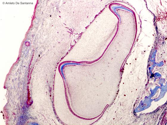
Figure E199. Human fetal mandible. Tooth bud inside the fetal mandible (lower jaw). H&E X25
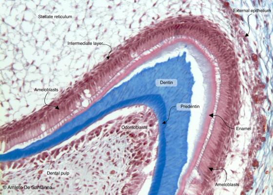
Figure E200. Human fetal tooth bud. Longitudinal section. Mallory-Azan X100
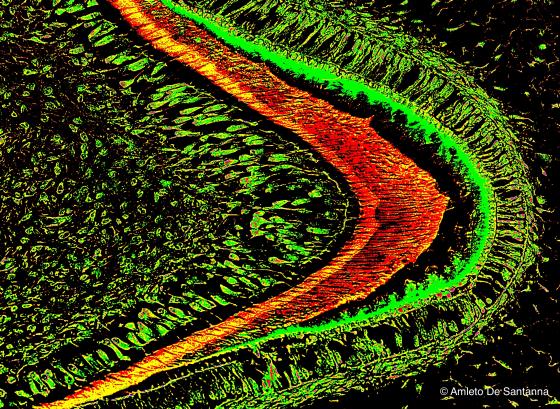
Figure E201. Human fetal tooth bud. Longitudinal section. This Mallory-Azan staining has been digitally processed to highlight the different structural elements. X100
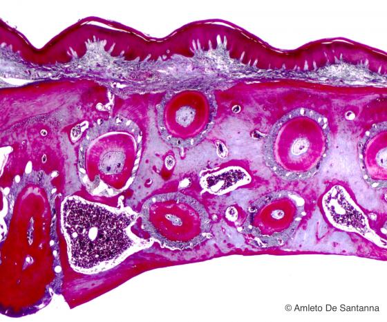
Figure E202. Dog hard palate. Transversal section at low magnification. Mallory-Azan X25
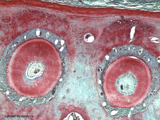
Figure E203. Dog hard palate. Palate transversal section where you can see two teeth cut transversely inside the ossifying mandible. Mallory-Azan X40
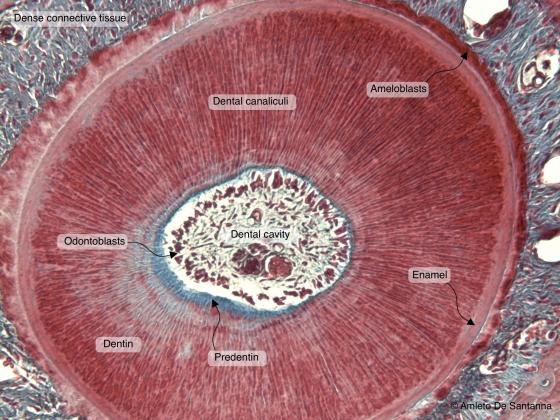
Figure E204. Dog tooth. Transversal section at higher magnification. You can easily see the enamel. Mallory-Azan X100
Crystalline lens
It originates from an ectoderm invagination: the optic cup. This is an elastic, transparent, oval, avascular, biconvex organ (presenting two poles and a larger central part called equator). It is positioned between the vitreous body and the posterior chamber and its function is to converge the image on the retina. The lens has three principal components: an external capsule of elastic connective tissue (lens capsule), a simple cuboidal epithelium (subcapsular epithelium) and both cortical and nuclear fibers, placed in the central area and representing highly differentiated epithelial cells. Cortical lens fibers contain nuclei and organelles not present in nuclear fibers.
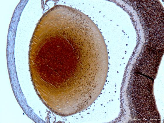
Figure E205. Mouse lens. Lens of a two days old mouse. Ignesti X40
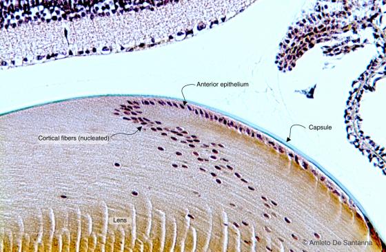
Figure E206. Mouse lens. Lens of a 28 days old mouse. This section shows the equator of a lens. You can see how the anterior epithelium (simple cuboidal) extends into the nucleated fibers (lens fibers), characteristic of this region of the lens. Ignesti X100
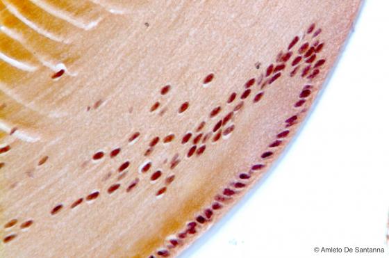
Figure E207. Mouse lens. Lens of a 42 days old mouse at high magnification. Ignesti X40
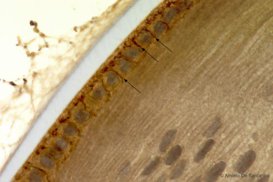
Figure E208. Mouse lens. Immunohistochemical reaction that highlights, by a brown precipitate, E-cadherins (arrows), transmembrane proteins that forms cell-cell adherent junctions in presence of calcium ions. Cadherins are the main proteins accountable of the maintaining of the bidimensional organization of the cells making up epithelia. There are different types of cadherins; E-cadherin is typical of epithelia. HRP X400
Nails
Nails are a derivative of epithelial tissue and their function is to protect the cutaneous tissue covering limb endings. Their structure is composed of two parts: the nail plate, a hard, external part consisting of several concentric layers of keratinized epithelial cells, and the nail bed, a deeper, highly vascularized part covered by a stratified squamous epithelium without stratum granulosum and stratum lucidum. At the base of the nail bed, there is a semicircular, lighter area, rich in air vesicles, called lunula. Its edge includes the nail matrix, a part of germinal epithelium that allows nail growth.
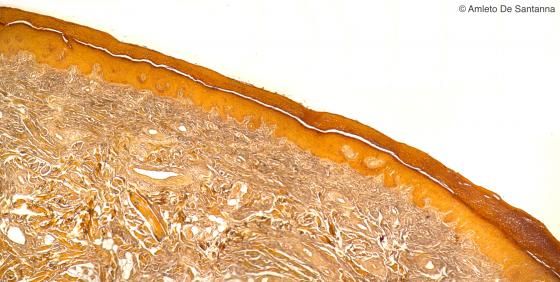
Figure E209. Human nail. Low magnification micrograph of a human nail where you can see the cornified stratifies squamous epithelium. Ignesti X25
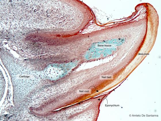
Figure E210. Human fetal nail. Mallory-Vannucci X63
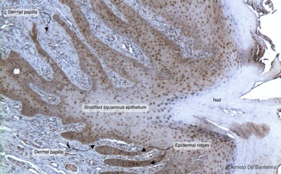
Figure E211. Human nail. The dermis forms papillae that enter in the epithelial tissue made of a stratified squamous epithelium where stratum granulosum and stratum lucidum are absent. Upon the epithelium there is the nail plate made of a thick layer of corneocytes. Hematoxylin DAB X40
Hairs
Hairs are a skin derivative. They present a free surface, the hair shaft, and a deeper part inside the epidermis, the hair root, whose base expands in the hair bulb. The hair shaft is composed of several layers of keratinized, elongated, squamous epithelial cells, rich in glycogen and organized in three concentric layers: the innermost part, or medulla, the intermediate, or cortex, and the outermost, or cuticle. Inside the cuticle, cells generally lose their nuclei and overlap like roof tiles. The cells composing the matrix are located on the same level as the hair bulb; they allow hair growth and, through differentiation, develop its numerous layers.
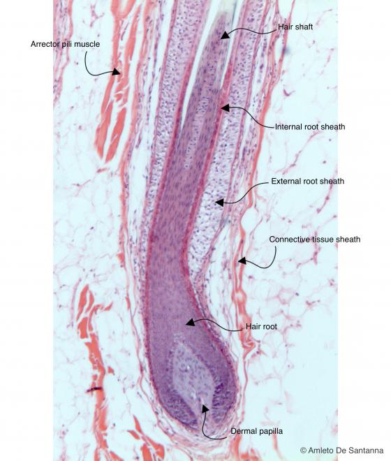
Figure E212. Human scalp. Hair follicle cut longitudinally. H&E X40
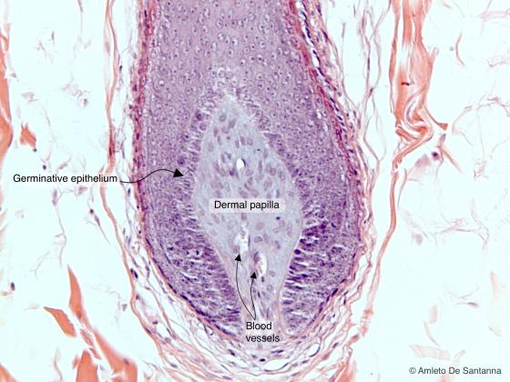
Figure E213. Bulb of a human hair follicle at high magnification. H&E X100
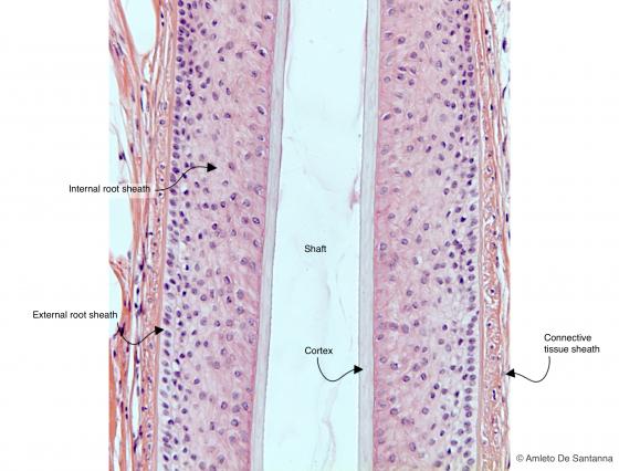
Figure E214. Human scalp. Longitudinal section of a hair. H&E X100
Other examples of highly differentiated epithelia
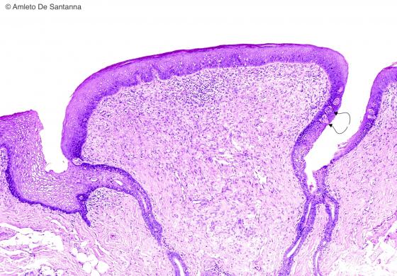
Figure E215. Human tongue. Circumvallate papilla lined by stratified squamous epithelium. You can see the taste buds (arrows). H&E X63
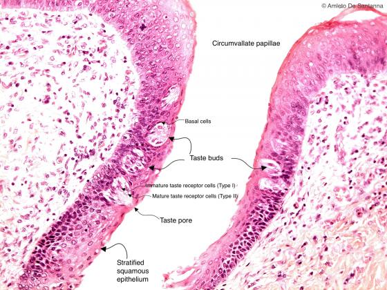
Figure E216. Human tongue at higher magnification. You can see the taste buds. Taste buds are made of three different cell types: mature neuroepithelial cells (type II cells, with pale cytoplasm and nucleus), immature neuroepithelial cells (type I cells, with elongated cytoplasm and dark nucleus) and basal cells, flattened against the basement membrane. H&E X200
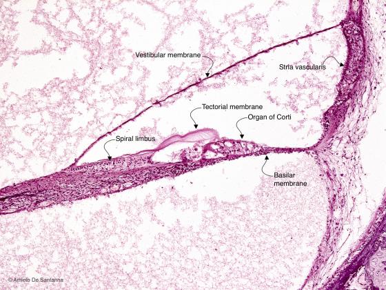
Figure E217. Human fetal inner ear. Transversal section of the coclear duct. H&E X100
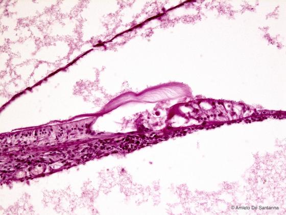
Figure E218. Human fetal inner ear at higher magnification. You can easily identify the organ of Corti. H&E X200