
Simple squamous epithelium
This epithelium is composed by a single layer of cells which are wider than they are tall. Their nucleus is flat and they are disposed in order to create a thin surface. This epithelium forms a thin, easily penetrable barrier, whose functions are to filter substances and exchange gasses. Here are some examples of simple squamous epithelium: the endothelium (that constitutes the tunica intima of blood vessels), the mesothelium (that constitutes serous membranes: pericardium, pleura and peritoneum), the epithelium constituting the renal capsule (Bowman’s capsule) of the renal corpuscle and the epithelium constituting alveolar walls.
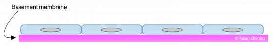
Figure E4. Schematic drawing of the simple squamous epithelium.
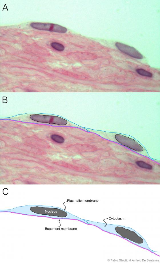
Figure E5. Digitally annotated micrograph of simple squamous epithelium. You can see extremely flattened, stretched cells with a central lengthened nucleus. Cytoplasm can be found in small or large amount and it is outlined in panel B.
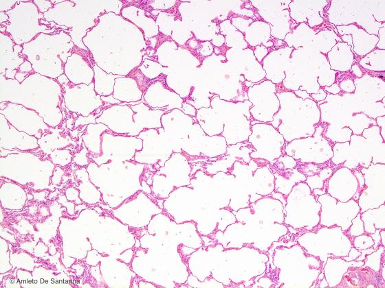
Figure E6. Rabbit lung. Pulmonar alveoli lined by simple squamous epithelium. Image obtained with the juxtaposition of two micrographs. H&E X63
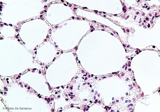
Figure E7. Rabbit lung. Pulmonar alveoli lined by simple squamous epithelium. H&E X200
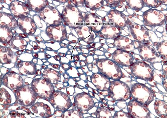
Figure E8. Rabbit kidney. Transversal section. In the central part of the micrograph, you can see the simple squamous epithelium that lines the descending limb of the loop of Henle. Mallory-Azan X100
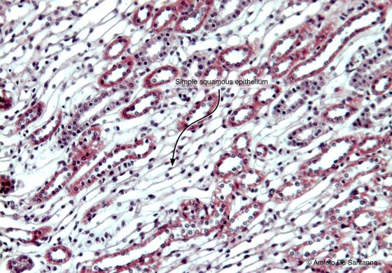
Figure E9. Rabbit kidney. Longitudinal section. You can easily see the simple squamous epithelium that lines the descending limb of the loop of Henle. H&E X100
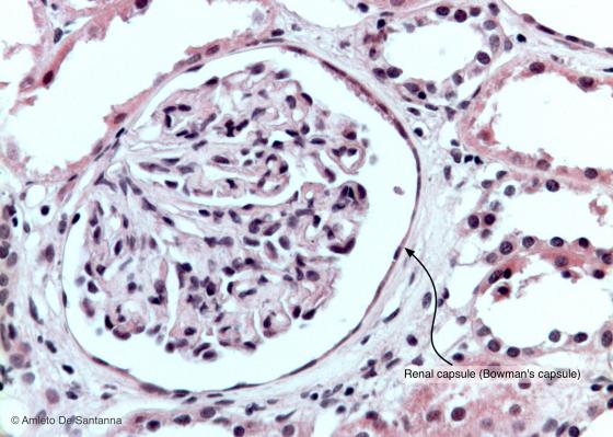
Figure E10. Mouse kidney. Simple squamous epithelium of the Bowmann capsule in the renal corpuscle. H&E X100
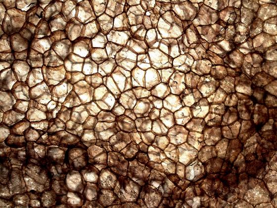
Figure E11. Rat omentum. Simple squamous epithelium obtained by sample stretching and stained with silver nitrate (view from above). X40
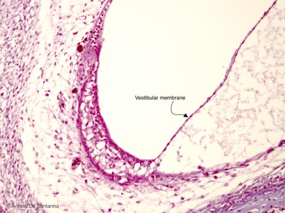
Figure E12. Human fetal internal ear. Vestibular (Reissner’s) membrane lined by simple squamous epithelium. H&E X40
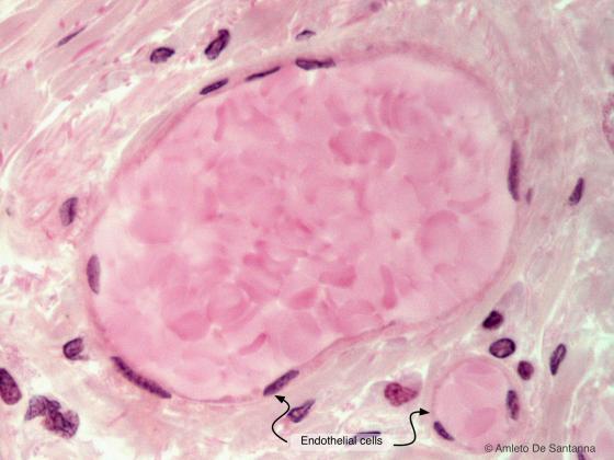
Figure E13. Human blood capillaries. High magnification micrograph that highlights the endothelial cells lining the capillaries. H&E X1000
Simple cuboidal epithelium
This epithelium is constituted by one cell layer. These cells are generally as tall as they are wide and present a round, central nucleus. When activated, they can take on an elongated shape, but they are distinguishable from the columnar epithelium in that their nucleus is always central. This epithelium mostly has a containment function: it covers the excretory ducts of exocrine glands, some tracts of the renal tubule and the thyroid follicles.
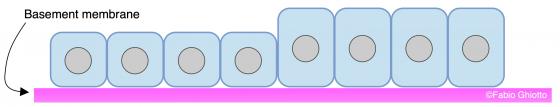
Figure E14. Schematic drawing of the simple cuboidal epithelium.
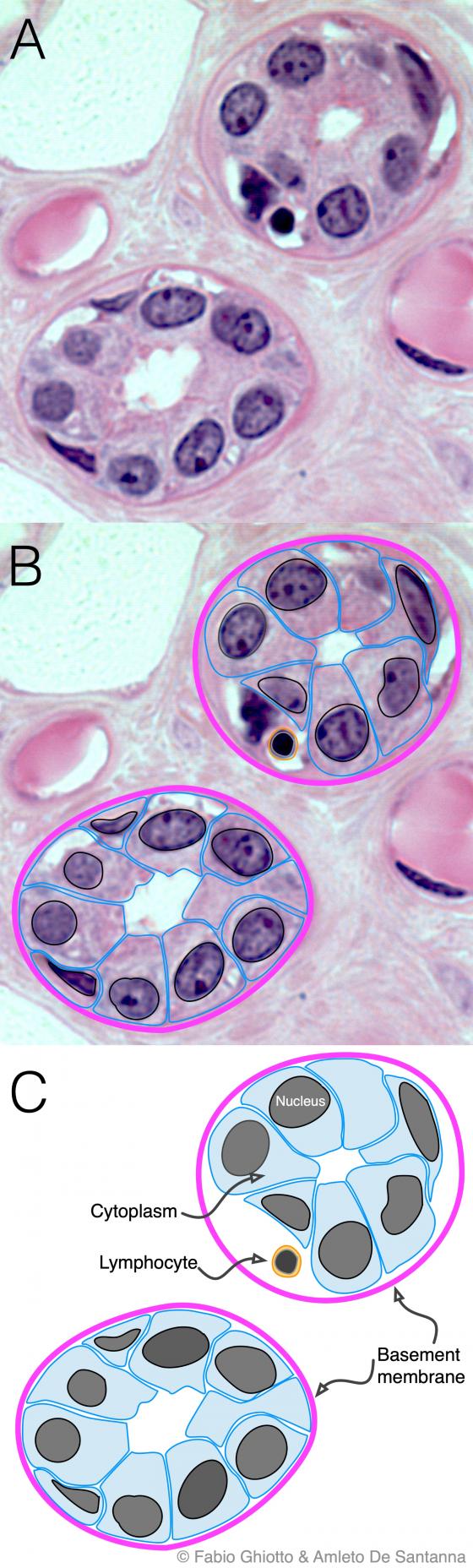
Figure E15. Digitally annotated micrograph of excretory ducts. The simple cuboidal epithelium is highlighted with a light blue outline. This epithelium is easy to recognize because it is formed by cells that are equally tall and large, and by the presence of a roundish nucleus always positioned in the center of the cell. Typically, the simple cuboidal epithelium lines ducts that contain fluids. H&E X400
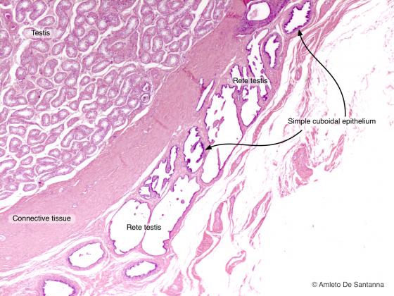
Figure E16. Rabbit testes. Rete testis. The canaliculi are lined by simple cuboidal epithelium. H&E X25
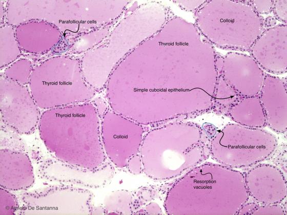
Figure E17A. Human thyroid. You can easily see the thyroid follicles lined by simple cuboidal epithelium and containing the colloid, the endocytotic vacuoles and the groups of parafollicular cells. Parafollicular cells (or C cells) can be recognized by an evident eosinophilic granulation in the cytoplasm. H&E X40
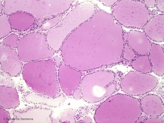
Figure E17B. Human thyroid. You can easily see the thyroid follicles lined by simple cuboidal epithelium and containing the colloid, the endocytotic vacuoles and the groups of parafollicular cells. Parafollicular cells (or C cells) can be recognized by an evident eosinophilic granulation in the cytoplasm. H&E X40
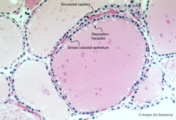
Figure E18. Human thyroid. Simple cuboidal epithelium that contributes to the formation of the follicle wall. H&E X100
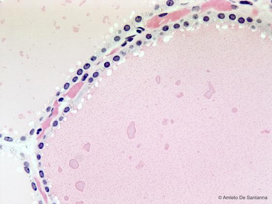
Figure E19. Human thyroid. Simple cuboidal epithelium at higher magnification. H&E X200
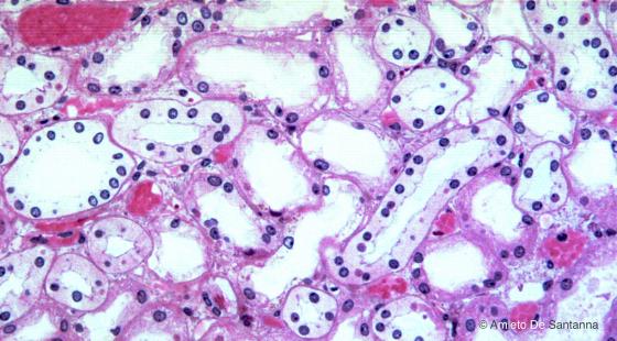
Figure E20. Human kidney. Renal tubules lined by simple cuboidal epithelium. H&E X100
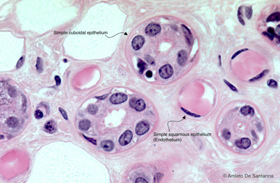
Figure E21. Human parotid gland. Secretory duct at high magnification lined by simple cuboidal epithelium. You can see two small blood vessels whose wall is lined internally by a layer of strongly flattened cells that form the endothelium of the vessels. H&E X40
Simple columnar epithelium
The cells forming this epithelium are much taller than they are large and are placed in the form of a palisade. Their nucleus is oval and placed in the first third of the cell, the nearest portion to the basal lamina. The simple columnar epithelium is very common and has multiple functions; this is why it can present various specializations on its surface. For example, in the gastrointestinal tract, it covers the luminal surface of the mucosa, and its cells present various microvilli on their apical surface; these are digitiform expansions of the cytoplasmic membrane, whose aim is to increase the absorbing surface and to ease the exchange of substances. Inside the salpinx, the simple columnar epithelium presents cilia because its function is no longer exchange, but that of creating amorphous currents which help the oocytes to enter the tubes, and descend (as a fecundated zygote) towards the uterus. At the same time, they impede the ascent of spermatozoa. The simple columnar epithelium can be found covering the excretory ducts of the renal tubule, without any known specialization.
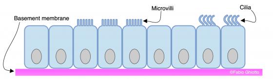
Figure E22. Schematic drawing of the simple columnar epithelium.
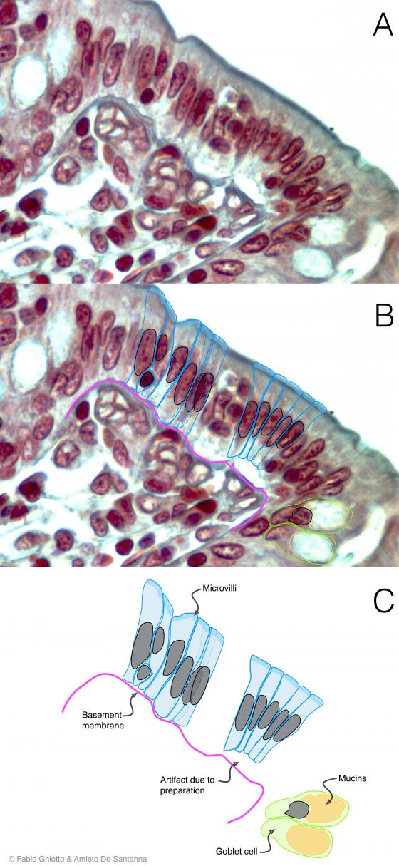
Figure E23. Digitally annotated micrograph of the columnar epithelium. The cells that make up this epithelium are taller than they are large and their nuclei, positioned in the lower third of the cytoplasm, are elongated. At the apex of the cells, you can see microvilli, finger-like protrusions that allow the increase of the exchange surface.
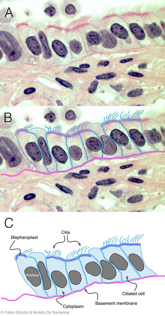
Figure E24. Digitally annotated micrograph of the ciliated columnar epithelium. The cells that make up this epithelium are taller than they are large. The nucleus of these cells, usually positioned in the lower third of the cell, is more elongated than the nucleus of cuboidal epithelium. In this particular example, you can see specialized structures, the cilia, located at the apex of the cells and that, with their beating activity, allow the movement of the fluid that covers the epithelium.
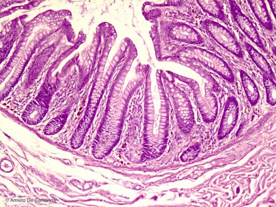
Figure E25. Human small intestine. Intestinal villi (lined by simple columnar epithelium with microvilli and goblet cells) and intestinal glands (cripts of Lieberkühn) are visible. H&E X40
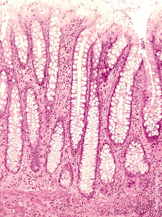
Figure E26. Human colon at high magnification. Simple columnar epithelium with goblet cells. H&E X200
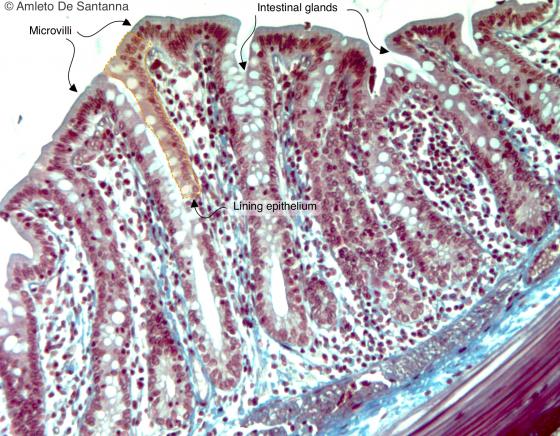
Figure E27. Rat colon. In the intestinal wall, you can see the folded mucosa lined by a simple columnar epithelium with several goblet cells. The columnar cells that form the epithelium are very tall and the nucleus, positioned at the base of the cell, is elongated. Mallory-Azan X400
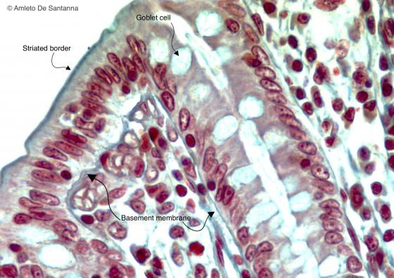
Figure E28. Rat colon. Simple columnar epithelium at high magnification. You can easily see the brush border, light blue on the surface of the columnar cells, and many goblet cells. You can also see the basement membrane. Mallory-AzanX400
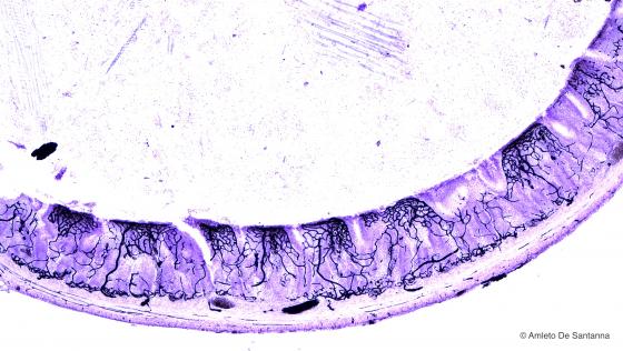
Figure E29. Rat intestine. Vital staining with India ink that shows the blood circulation in the intestinal wall. India ink X40
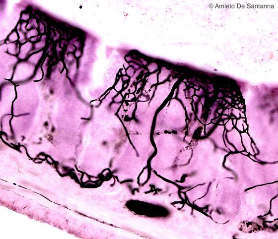
Figure E30. Rat intestine. High magnification of the intestinal wall stained with India ink. India ink X200
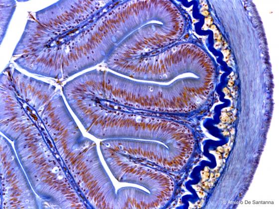
Figure E31. Fish intestine. Simple columnar epithelium of the intestinal mucosa. In the submucosa is present a thick layer of elastic connective tissue, stained deep blue, that prevents a possible perforation or breakage of the intestinal wall. Modified Mallory method X100
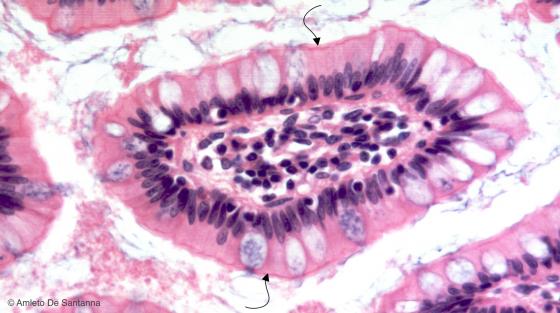
Figure E32. Human ileum. Transverse section of intestinal villi where you can see the simple columnar epithelium and the brush border (arrows). H&E X200
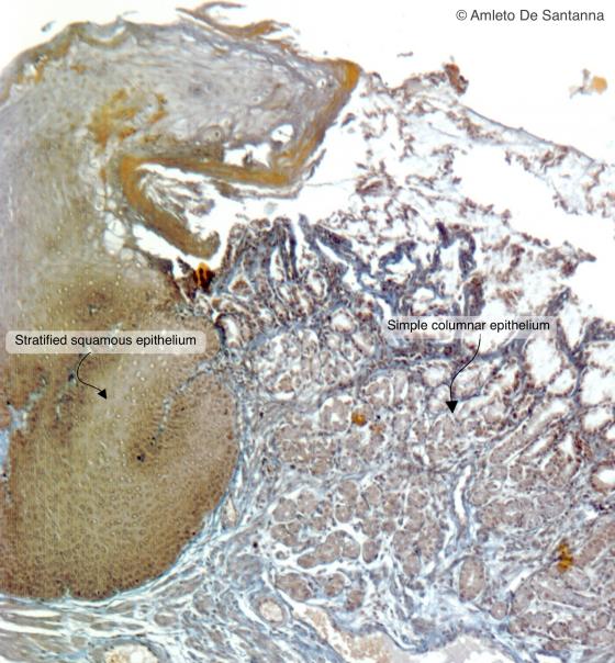
Figure E33. Rabbit gastro-esophageal junction. The transition from the stratified squamous epithelium of the esophagus to the simple columnar epithelium of the stomach is visible. Mallory trichrome staining X63
Figure E34. Human gallbladder. Low magnification. You can see the ciliated simple columnar epithelium that lines the gallbladder (arrows). Mallory-Azan X40
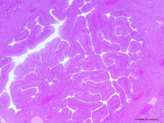
Figure E35. Human oviduct. The oviduct is lined by ciliated simple columnar epithelium. H&E X40
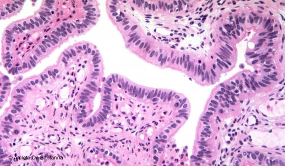
Figure E36. Human oviduct. Micrograph at higher magnification where you can see the cilia of the epithelium. H&E X100
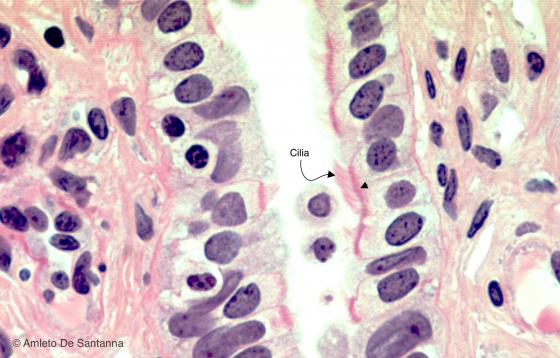
Figure E37. Human oviduct. In this high magnification micrograph, you can see the cilia of the apical domain of the simple columnar epithelium that lines the oviduct. The dark pink line at the base of the cilia is the specific structural system that anchors the cilia to the cell (arrowhead). H&E X400
Pseudostratified epithelium
In this epithelium, every single cell is in direct contact with the basal lamina, but only some of them can reach the surface. In tissue specimens, the nuclei are found on different levels; they are staggered, but never overlapped. The cells constituting this epithelium may present various shapes. They have a cytoplasmic bulge that contains the nucleus. This bulge can be placed either on the top or on the bottom of the cell. Thanks to this distribution, when analyzed using the microscope, this epithelium erroneously looks like a stratified epithelium (hence the adjective “pseudostratified”). There are two types of pseudostratified epithelium: the pseudostratified ciliated epithelium and the pseudostratified epithelium with stereocilia.
The function of the pseudostratified ciliated epithelium is mostly to purify the inspired air from dust and environmental pathogens. Goblet cells secrete mucus (made of highly glycosylated and hydrated glycoproteins) that traps impurities. Subsequently, mucus is evenly spread and moved out by the upper ciliated cells. The same process takes place in the trachea, where mucus is moved upwards (mucociliary elevator). This epithelium is to be found in the majority of respiratory tracts, the auditory tube (Eustachian tube), in some parts of the tympanic cavity and lacrimal sac.
The pseudostratified epithelium with stereocilia is found in the epididymis. The cell surface of this epithelium presents “tuft” extensions, the stereocilia, whose aim is to collect and nourish scattered spermatozoa in the lumen of the epididymis.

Figure E38. Schematic drawing of the pseudostratified epithelium.
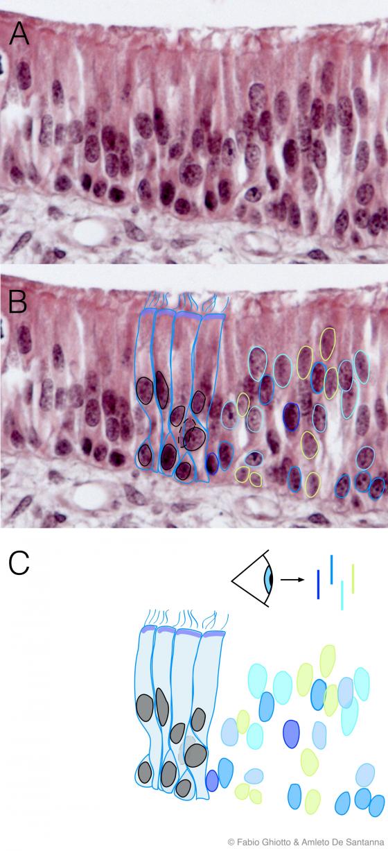
Figure E39. Digitally annotated micrograph of the pseudostratified epithelium. This epithelium is made up by tall cells that reach the free surface and by lower cells (staminal cells) that do not reach the free surface. This cell organization results in cell nuclei positioned at different heights and giving the impression you are looking at a stratified epithelium (hence the name pseudostratified). The different colors show nuclei belonging to cells in different planes (blue shows the longitudinal plane nearest the observer, light green the farthest).
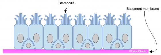
Figure E40. Schematic drawing of the pseudostratified epithelium with stereocilia.
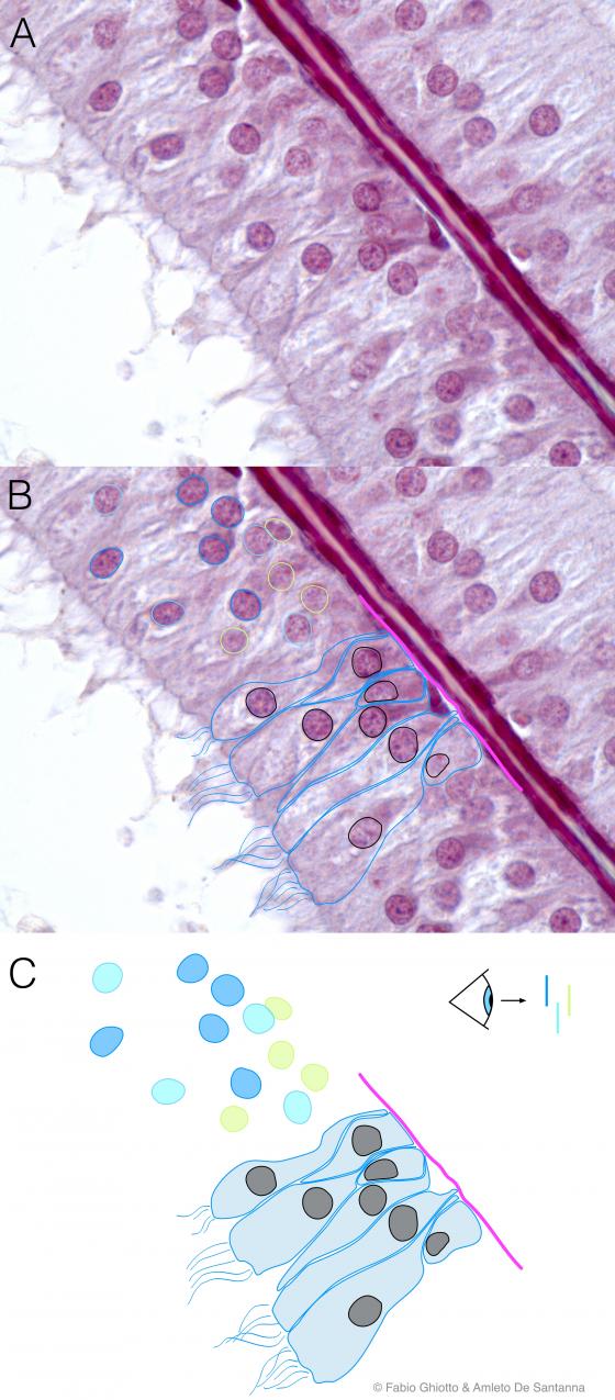
Figure E41. Digitally annotated micrograph of pseudostratified epithelium with stereocilia.This epithelium is structurally similar to the pseudostratified epithelium with cilia but, having a different function, it has stereocilia, formations that are structurally like microvilli. Stereocilia have two different functions: they need to be long enough to follow amorphous corrents to group together the spermatozoa; and they have an exchange function with the cells present in the epidymis. The different colors show nuclei belonging to cells in different planes (blue shows the longitudinal plane nearest the observer, light green the farthest).
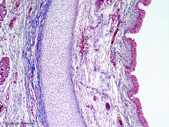
Figure E42. Human trachea. The trachea is lined by pseudostratified epithelium with cilia (also called respiratory epithelium). Mallory-Azan X25
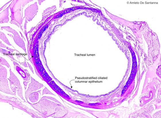
Figure E43. Mouse trachea. Histochemical staining of the trachea that shows acid glycosaminoglycans in blue (alcian blue) and neutral ones in magenta (PAS). Alcian-PAS X25
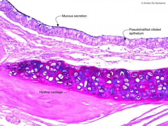
Figure E44. Mouse trachea. Histochemical staining of the trachea at higher magnification that shows acid glycosaminoglycans in blue (alcian blue) and neutral ones in magenta (PAS). Alcian-PAS X200
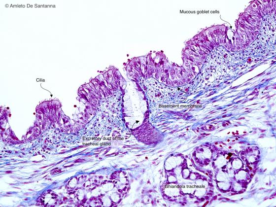
Figure E45. Human trachea. Pseudostratified epithelium. On the surface of the cells you can see the cilia whose function is to create currents that allow the movement of mucus on the surface of the epithelium. Azan-Mallory X100
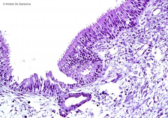
Figure E46. Human trachea. Epithelium of the trachea at higher magnification. H&E X200
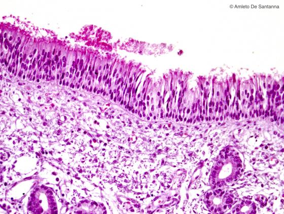
Figure E47. Human fetal nasal cavities. Ciliated pseudostratified epithelium. H&E X100
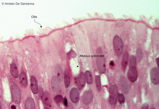
Figure E48. Human fetal nasal cavities. Ciliated pseudostratified epithelium. H&E X400
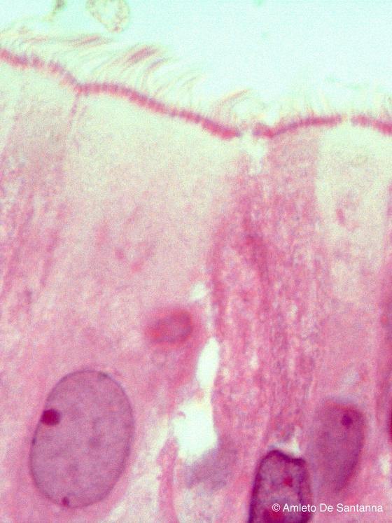
Figure E49. Human fetal nasal cavities. Ciliated pseudostratified epithelium at higher magnification. You can easily see the cilia and their anchoring system. H&E X1000
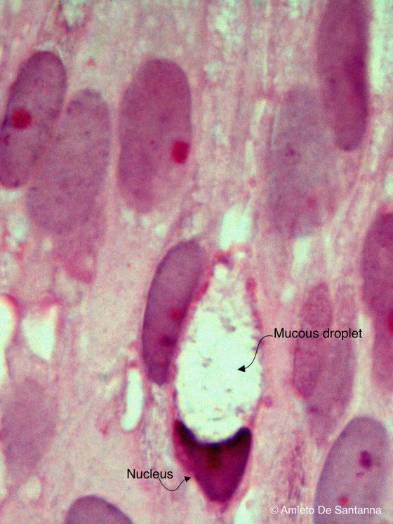
Figure E50. Human fetal nasal cavities. Ciliated pseudostratified epithelium at higher magnification. You can see a mucous cell. H&E X1000
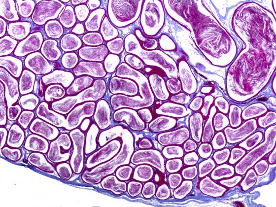
Figure E51. Rat epididymis. Low magnification micrograph where you can see the spermatozoa in the head of the epididymis. Azan-Mallory X25
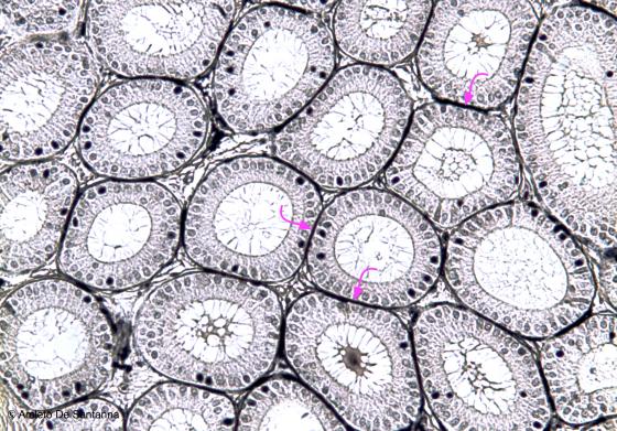
Figure E52. Mouse epididymis. Pseudostratified epithelium with stereocilia of the epididymis. You can see the basal membrane stained black (arrows). Elective staining for cell borders. Iron Hematoxilyn X100
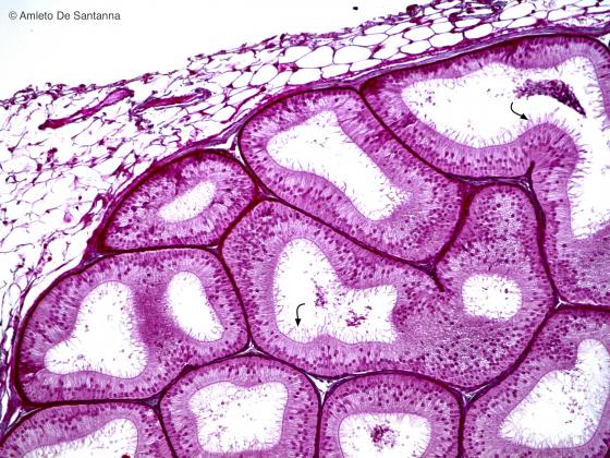
Figure E53. Rabbit epididymis. Pseudostratified epithelium with stereocilia (arrows). Stereocilia are not real cilia but long and irregular microvilli. Their function, that is to nourish and to group together the spermatozoa, explain their length and peculiar form. Mallory-Azan X100
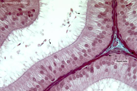
Figure E54. Rabbit epididymis. Pseudostratified epithelium with stereocilia at high magnification. You can see the stereocilia, long cytoplasmic filaments on the apical domain of the cells, and a layer of squamous or cubical cells close to the basement membrane called basal cells. Mallory-Azan X200
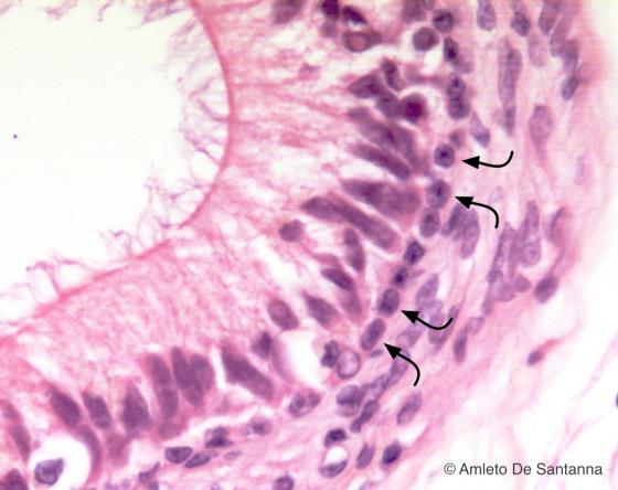
Figure E55. Human epididymis. Pseudostratified epithelium with stereocilia. You can see the basal cells close to the basement membrane (arrows). H&E X200
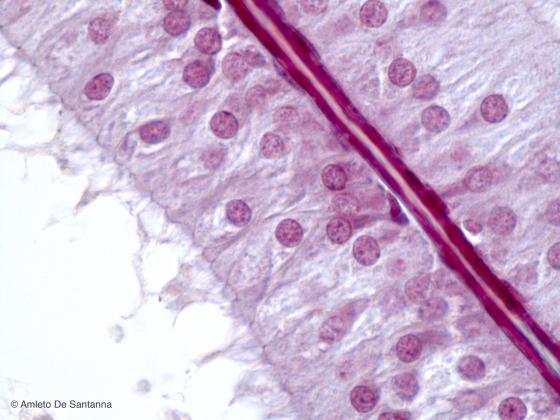
Figure E56. Rabbit epididymis. High magnification micrograph where you can easily see the pseudostratified epithelium with stereocilia. The basement membrane is stained bright red. Mallory-Azan X250
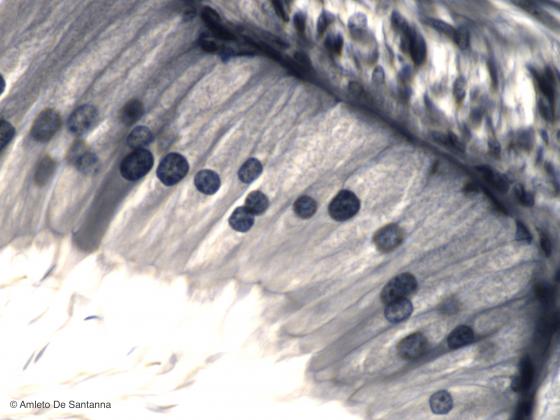
Figure E57. Mouse epididymis at high magnification stained with a selective staining that highlight cellular borders and stereocilia. Iron Hematoxylin X300
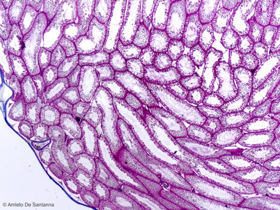
Figure E58. Rat testis. Low magnification micrograph where you can see the seminiferous tubules of the testis. Mallory-Azan X25