
As immunohistochemistry, immunofluorescence is a highly specific method traditionally used to reveal certain antigens present in the tissue or in the cells to be analyzed. A specific antibody must be placed on the section of tissue or cells properly prepared. If this antibody is directly bound to fluorescent molecules (fluorochromes, e.g. FITC, TRITC, PE, etc.), the staining technique is called ‘direct immunofluorescence’. Instead, if this antibody is further recognized by a secondary antibody which is conjugated to the fluorochrome, then the staining technique is called ‘indirect immunofluorescence’. The samples “stained” by both types of immunofluorescence will be then analyzed using a fluorescence or a confocal microscope.
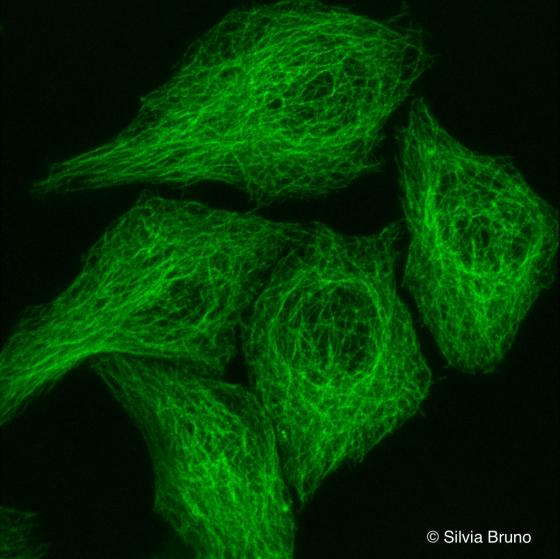
Figure S34. HeLa cell line. In this confocal microscopy micrograph, the cytoskeletal component made of tubulin has been highlighted with a specific antibody. The image is a three-dimensional reconstruction obtained using serial planes. Courtesy of Silvia Bruno, Department of Experimental Medicine, University of Genoa
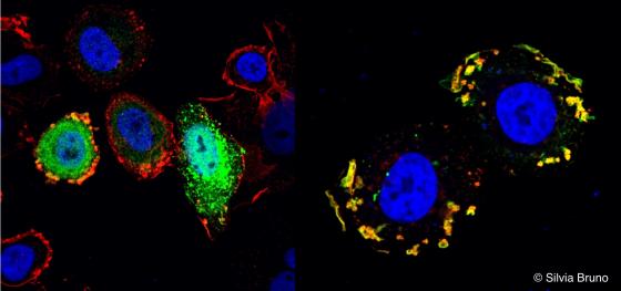
Figure S35. (A) SK-BR-3 cell line transfected with a FLAG-SH3BGRL3 construct. The protein SH3BGRL3 has been revealed using a green fluorescent anti-FLAG antibody, while the red fluorescence is due to the use of an anti-Erbb2 antibody. The nucleus has been stained blue with DAPI. (B) The SK-BR-3 cell line has been stained with anti-Erbb2 (red) and anti-myosin 1C (green) antibodies. The nucleus has been stained with DAPI (blue). Courtesy of Silvia Bruno, Department of Experimental Medicine, University of Genoa.
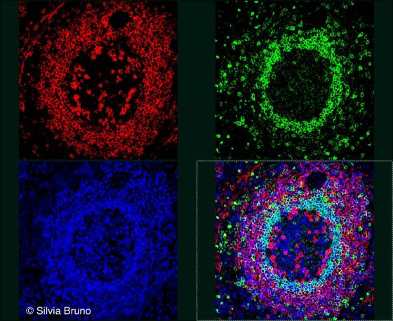
Figure S36. Human spleen. Secondary lymphoid follicle. The nuclei of all the cells of the follicle have been stained blue with DAPI. The B lymphocytes of the follicle have been stained with anti-CD27 (red) and anti-IgD (green) antibodies. In the lower quadrant on the right, different colors superimpose: B cells expressing CD27 only are pink (a virtual staining for blue+red), B cells expressing IgD only are light blue (blue+green). In this follicle, there are no double positive B cells (for both CD27 and IgD). Courtesy of Silvia Bruno, Department of Experimental Medicine, University of Genoa.
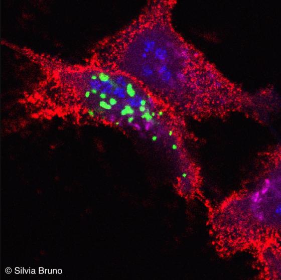
Figure S37. HeLa cells transfected with PML-GFP fusion protein construct and stained with anti-fibrillarin (blue), anti-GM130 (pink) and anti-MHC class I(red) antibodies. Fibrillarin is a protein found in the nucleolus, GM-130 a protein in the cis-Golgi and class I MHC molecules in the plasmatic membrane. Courtesy of Silvia Bruno, Department of Experimental Medicine, University of Genoa.