
The epithelial tissue is composed by morphologically differentiated cells which are tightly connected by specialized junctions: desmosomes, tight junctions and gap junctions. Therefore, they present minimum extracellular matrix.
The morphological and spatial organization of epithelia allows them to accomplish various types of tasks such as protection, exchange, lubrication of external and inner surfaces.
These tasks are accomplished by highly specialized cells, which are employed in different activities such as absorption, secretion, transport, gas exchange, sliding (of two surfaces) and sensory functions. This is the reason why the epithelial tissue is so differentiated: it depends on the specific function fulfilled.
Each epithelium presents a basement membrane composed by proteoglycans (PAS positive) which separates the epithelium from the underneath connective tissue. Beneath the basement membrane, we find loose reticular connective tissue which contains type III collagen. This membrane can be easily highlighted by the periodic acid-Schiff’s reagent staining method (PAS staining method).
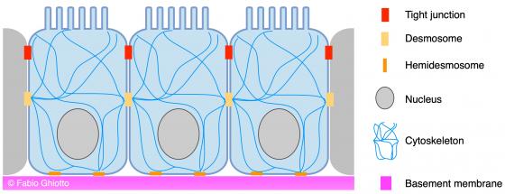
Figure E1. Schematic drawing showing the organization of the lining epithelium
Epithelial tissue derives from the three germ layers; for example, the epidermis and the corneal epithelium derives from the ectoderm. The mesoderm gives rise to the kidney epithelium and to both female and male genital organs epithelium. Gastric glands, intestinal glands, liver, pancreas, the lining of blood vessels (known as endothelium) and of the serous cavities (known as mesothelium) derive from the endoderm.
Covering and lining epithelia
Regardless of the shape of its cells, a covering or lining epithelium can assume two different appearances: on the one hand, it can look like a unique layer of joined cells leaning on a thin basal lamina. In this case, we refer to simple epithelium. On the other hand, it can be composed by two or more overlapping cell layers, in which only the deepest layer leans on the basal lamina. In this case, we talk about stratified epithelium.
The cells composing the epithelial tissue are polarized, this means that their surface can accomplish different and specific functions. More specifically, epithelial cells present an apical and a basolateral domain. The apical domain establishes the functional features of the epithelium. It can either present features which makes liquids flow on the surface of the epithelium itself (vibrating cilia) or increase the cell surface by means of specific devices (microvilli and stereocilia).
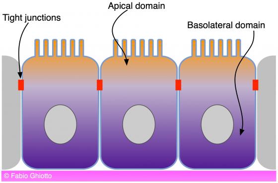
Figure E2. Schematic drawing showing the apical and the basolateral domains of the lining epithelium.
Covering and lining epithelia are classified according to:
- The number of cell layers
- The cell shapes
- The presence and type of specialized features on the apical domain.
In stratified epithelia, the categorization is carried out by considering only the shape of the cells present in the more superficial layer, since the deeper ones are usually cubic or cylindrical.
The following epithelia are simple:
- The simple squamous epithelium
- The simple cuboidal epithelium
- The simple columnar epithelium
- The pseudostratified epithelium
The following epithelia are stratified:
- The stratified squamous epithelium
- The stratified cuboidal epithelium
- The stratified columnar epithelium
- The transitional epithelium
Glandular epithelia
The epithelial tissue whose main function is secretion is known as glandular epithelium.
This specific type of tissue is defined exocrine when the substance is secreted out in a body cavity or outside the body by means of the excretory ducts, while it is known as endocrine in case the substance is released in blood circulation.
Highly specialized epithelia
In order to accomplish specific functions, these epithelia have differentiated their morphological and functional features. In this way, such features have become extremely different from those of the “normal” epithelial tissue: for this reason, we have decided to dedicate to these tissues a separate analysis. In this category, we can find the crystalline fibers, the tooth enamel and the cutaneous annexes such as hair, body hair and nails.
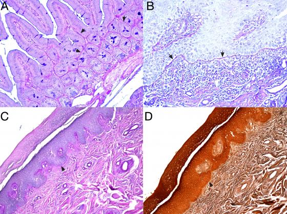
Figure E3. Panels A and B. The basement membrane, stained magenta with PAS staining (arrows), is clearly visible in a section of rat small intestine (panel A) and of human nail (panel B). Panels C and D compare adjacent sections of human nail, one stained with the PAS method (panel C) and one with the Ignesti’s method (panel D). It is obvious how the PAS staining makes visible the basement membrane, while the Ignesti’s staining does not.