
This tissue is specialized in creating movement through the contraction of its cells. If its function changes, then also its morphology and structure does. There are three types of muscle tissue:
- Striated skeletal muscle
- Striated cardiac muscle
- Smooth muscle
Striated skeletal muscle
Striated skeletal muscle is formed during embryonic life, from the fusion of several myoblasts. In this way, a myotube is created; this means that cells form a cellular syncytium with a cylindrical, elongated shape, called muscle fiber. The single muscle fiber is characterized by transverse stripes forming the typical bands, either lighter or darker, visible with a light microscope. The nuclei are extremely elongated and peripheral. A thin layer of reticular connective tissue, called endomysium, surrounds each fiber. Fibers are organized in fascicles surrounded by connective tissue, called perimysium. These fascicles are in turn organized to form a single muscle, which is surrounded by connective tissue that forms the epimysium. This arrangement allows the striated muscle to contract (shorten) and relax (lengthen) quickly, with no damage. The contraction of this type of muscle is voluntary.
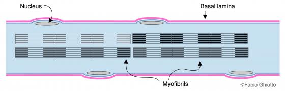
Figure M1. Schematic drawing of a striated muscle fiber.
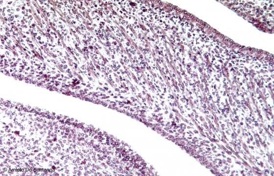
Figure M2. Mouse embryo. Tongue. Rhabdomyoblasts are cells of mesenchymal origin that, after a fusion and differentiation process, originate striated muscular fibers. H&E X100
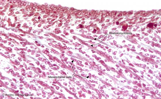
Figure M3. Mouse embryo. Tongue at higher magnification. You can see the morphological differences between the rhabdomyoblasts, small and elongated cells with well-stained cytoplasm that are the precursors of the muscular cells, and the mesenchymal cells, not yet differentiated, that are roundish and with a poorly stained cytoplasm. Mallory-Azan X200
Figure M4. Human tongue. Striated muscle fibers. The striated muscle is characterized by bands, dark bands (A bands) and light bands (I bands), easily seen in longitudinal section. Moreover, the fibers have elongated thin nuclei, positioned peripherally, and form small bundles partitioned by thin connective sheets. H&E X100
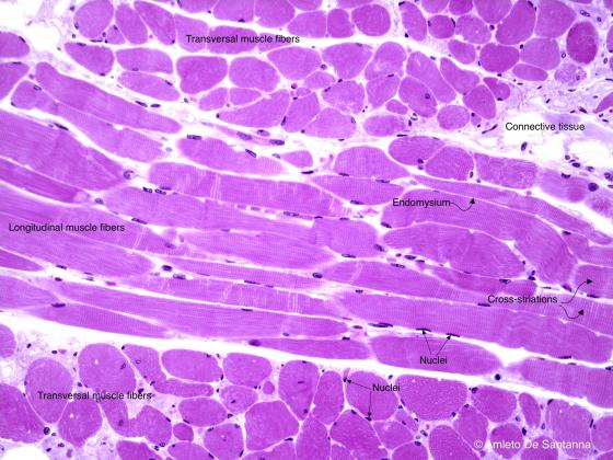
Figure M5. Human tongue. Striated muscle fibers. You can see the bands, the elongated and peripherally positioned nuclei, and the mesh of reticular connective tissue that surrounds each muscle fiber (endomysium). H&E X200
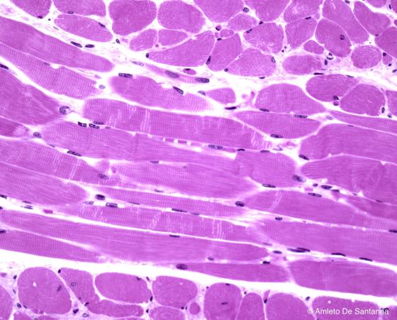
Figure M6. Human tongue. Striated muscle fibers at higher magnification. H&E X250
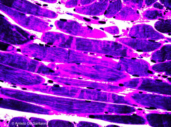
Figure M7. Human tongue. Striated muscle fibers. Digitally processed micrograph in order to highlight the reticular connective tissue (pink) that surrounds each muscle fiber bundle (purple). X250
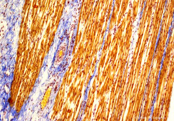
Figure M8. Human tongue. Striated muscle fibers. This staining allows to distinguish clearly the muscle fibers (ocher) and the connective tissue (light blue). Ignesti X100
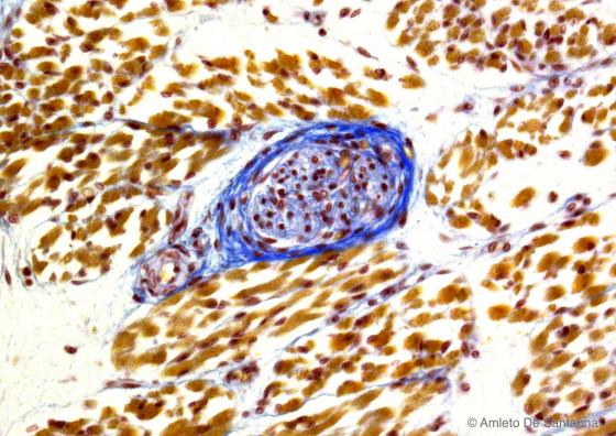
Figure M9. Human fetal tongue. Muscle spindle (blue) enclosed in striated muscle tissue. Ignesti X250
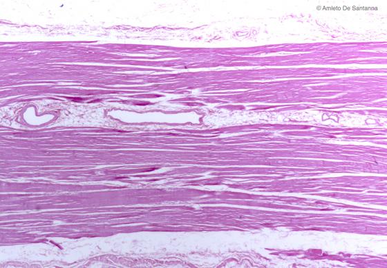
Figure M10. Human diaphragmatic muscle. H&E X20
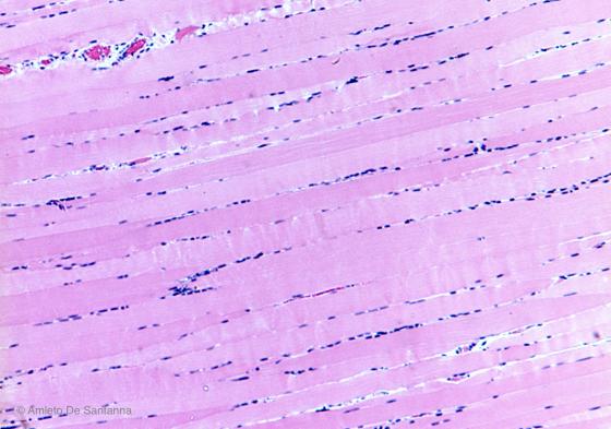
Figure M11. Human striated skeletal muscle. You can see the parallel alignment of the striated muscle fibers and the peripheral location of the nuclei. H&E X40
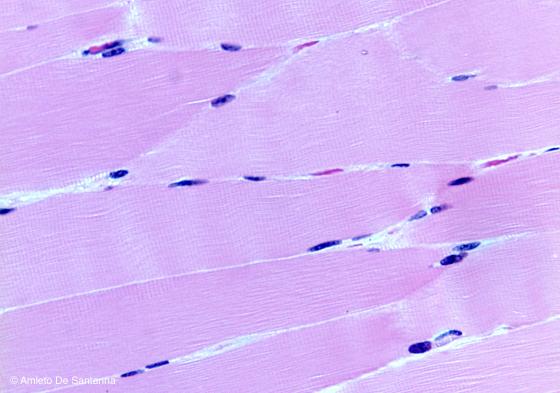
Figure M12. Human striated skeletal muscle at higher magnification. Semithin section in which you can recognize the thin connective sheets that surround each muscle fiber (endomysium), the strongly stretched out and peripherally positioned nuclei and the cross-banding pattern. H&E X200
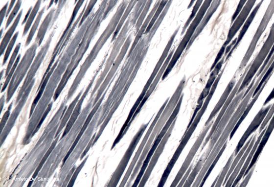
Figure M13. Human striated skeletal muscle. You can see the organization of the tissue in small bundles. Iron hematoxylin X63
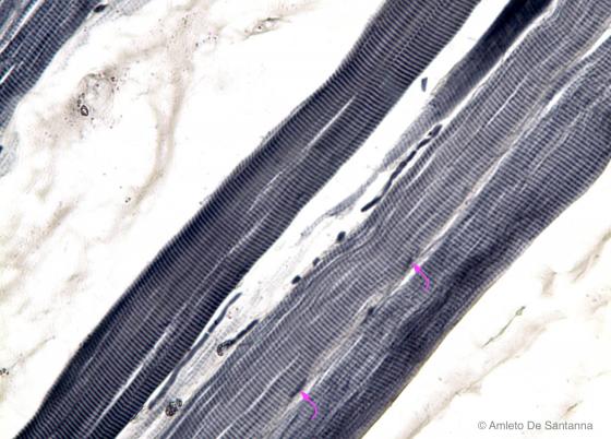
Figure M14. Human striated skeletal muscle at higher magnification. Staining that is selective for the cell borders and for the characteristic bands of the striated muscle. You can also see a few nuclei, elongated in shape and positioned peripherally (arrows). Iron hematoxylin X200
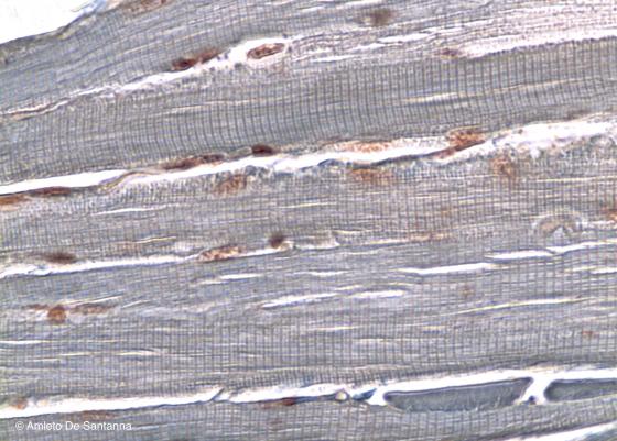
Figure M15. Rabbit tongue. Striated skeletal muscle tissue at higher magnification. Also with this staining, you can appreciate, in longitudinal section, the characteristic cross-banding of the striated muscle tissue. Ignesti X200
Striated cardiac muscle
The cells composing the striated cardiac muscle are named cardiomyocytes; they have a cylindrical, shape (often with a bifurcation) and are 85-100 µm long and 15 µm wide. Cardiomyocytes present highly visible transverse stripes (like the striated skeletal muscle) and only one, very evident, central nucleus (like smooth muscle). A particular feature of this type of muscle is the presence of intercalated discs, which are specific devices that connect the cells. In such discs, there are gap junctions that allow electrical coupling among myocardial cells. In this way, they enable a simultaneous contraction.
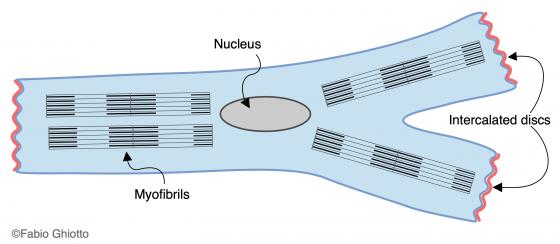
Figure M16. Schematic drawing of a cardiomyocyte.
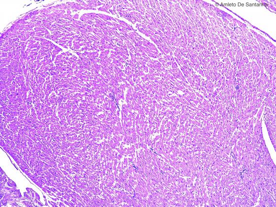
Figure M17. Human cardiac muscle tissue. An overview. Notwithstanding the low magnification, you can see some characteristics that are typical of this tissue: the cardiac muscle cell has an irregular shape and a centrally positioned nucleus. H&E X40

Figure M18. Human cardiac muscle tissue at higher magnification. You can easily see the cardiac muscle cells with their central nuclei. The cardiac muscle cells, after a short straight course, divide in two, resembling a Y shape, and connect with the adjoining cardiac muscle cells (arrows). With this staining, the cross-banding is not easily appreciable. H&E X200
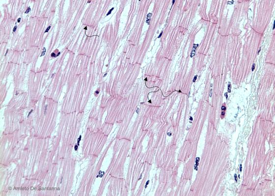
Figure M19. Human cardiac muscle tissue. Thicker section of cardiac muscle in which you can easily see both the cross-banding and the intercalated discs that function as cell to cell junction system (arrows). H&E X100
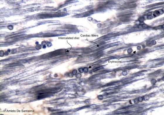
Figure M20. Human cardiac muscle tissue at high magnification. At the center of the image, an intercalated disc is easily seen. The roundish structures clearly seen in the specimen are red blood cells inside the numerous capillary vessels of the heart. Iron hematoxylin X400
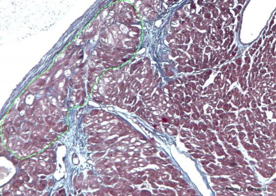
Figure M21. Human cardiac muscle tissue. The Purkinje fibers are large, globular, with one or two nuclei and rich in mitochondria and glycogen, are easily seen inside the area delimited by the dotted line. These characteristics make them, at the optical microscope, lighter and bigger that the regular cardiac muscle cells. Azan-Mallory X63
Smooth muscle
Smooth muscle is formed by single cells measuring from 20 µm to 0.5 mm in length and containing a very evident, ovoid, central nucleus. Smooth muscles are not voluntary and are controlled by the Visceral Nervous System (Autonomic Nervous System) and the Endocrine System. Smooth muscle presents two types of contraction: a rhythmic contraction with regular impulses that propagate throughout the tissue, and a “tonic” contraction that guarantees a condition of partial contraction called “muscle tone”. Depending on their function, smooth muscle cells display different spatial positions. Inside the vessels, they follow a round path. In this way, they are able to change the lumen after contracting. Whereas, in viscera and large hollow organs smooth muscle cells generally follow a round, inner path and an outer, longitudinal path, which is positioned orthogonally with respect to the first path. This arrangement allows a higher tightness of the viscera and facilitates the movement of its content, due to the peristaltic activity of the wall. In some hollow organs, such as the urinary bladder and uterus, muscle sheets follow a less regular path and create a dense meshwork. Therefore, it can be noticed that they have multiple functions: they support and allow muscle contraction and facilitate the spatial extension, increasing, in this way, the containment capacity of the organ itself. This arrangement of the muscle tissue is defined “plexiform”.
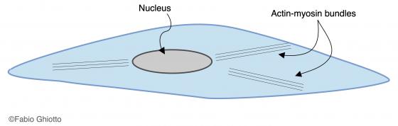
Figure M22. Schematic drawing of a smooth muscle cell.
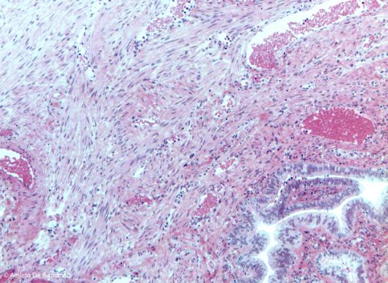
Figure M23. Human fallopian tube. Plexiform smooth muscle of the fallopian tube composed of elongated cells with a central nucleus and organized without an apparent order. H&E X63
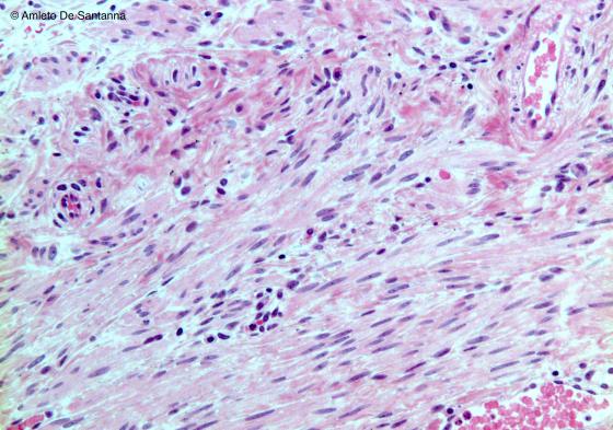
Figure M24. Human fallopian tube at higher magnification. You can easily see the specific characteristics of the smooth muscle cells: they are elongated, with and oval central nucleus easy to appreciate (both in longitudinal and transverse section), are not organized in bundles and lack the cross-banding. H&E X100
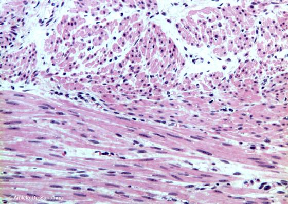
Figure M25. Muscular tunica of human intestine. Smooth muscle cells in both longitudinal and transverse section. In both cases, the nuclei are central to the cells. The arrangement in two muscle layers, one circular and one longitudinal, is typical of the muscular tunica of the majority of the hollow organs. H&E X100

Figure M26A. Mouse intestine in transversal section. The two layers of muscle tissue, circular inside and longitudinal outside, are easily seen. This arrangement in suitable for the peristaltic movements the intestine performs. Mallory trichrome staining X100
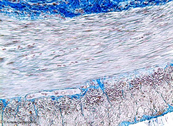
Figure M26B. Mouse intestine in transversal section. The two layers of muscle tissue, circular inside and longitudinal outside, are easily seen. This arrangement in suitable for the peristaltic movements the intestine performs. Mallory trichrome staining X100
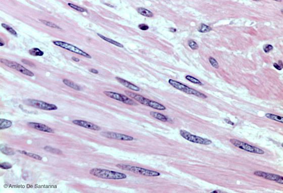
Figure M27. Muscular tunica of human intestine at high magnification. You can see the longitudinal bundles of smooth muscle cells, the centrally positioned nuclei and the lack of cross-banding. H&E X400
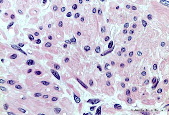
Figure M28. Muscular tunica of human intestine. Transversal bundles of smooth muscle cells. In the muscle cells, the centrally positioned nuclei are easily seen. H&E X400
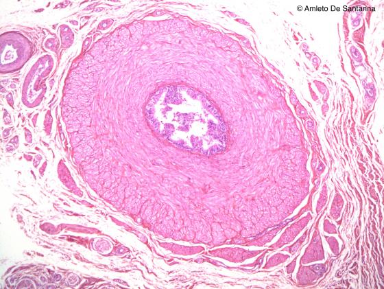
Figure M29. Human deferens duct. Smooth muscle. H&E X40
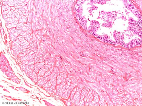
Figure M30. Human deferens duct. Smooth muscle. The different orientation of the smooth muscle cells, circular and longitudinal, are easily seen. H&E X100
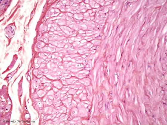
Figure M31. Human deferens duct at higher magnification. H&E X200