
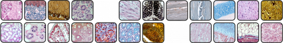
The aim of this Histology Atlas is to provide students with a concise explanation of the different staining methods used for histological specimen preparations and of the main characteristics of the different tissues. The text is accompanied by extensive iconographic documentation (501 images) illustrating the concepts covered by the Atlas.
Important note
All the pictures found in this Atlas refer to samples obtained from histological findings and preparations belonging to the Histology Section of the Department of Experimental Medicine at the University of Genoa. Histological samples of human fetus and pets, such as dogs, come from the archives of the Section, where they have been stored for more than 50 years. Samples related to marine mammals come from stranded specimens. Such samples have been used in scientific studies in collaboration with the Department of Life and Environmental Earth Sciences (DISTAV) at the University of Genoa.
This Atlas of Histology was produced with contributions from:
Amleto De Santanna: histological samples, stainings and micrographs
Amleto De Santanna, Fabio Ghiotto and Alessandro Moretta: italian text
Fabio Ghiotto: drawings and graphics
Martina Santi: translation into English
Ilaria Rizzato: linguistic supervision
Fabio Ghiotto and Silvia Bruno: scientific supervision of the translation
Amleto De Santanna, Fabio Ghiotto, Silvia Bruno and Alessandro Moretta: Department of Experimental Medicine, Ilaria Rizzato: Department of Modern Languages and Cultures, University of Genoa
